
Introduction
Bleeding is the most frequent complication of peptic ulcer (PU), and this in turn represents the main cause of upper gastrointestinal bleeding (UGIB). The bleeding risk of a PU is stratified according to the Forrest classification during endoscopy. High-risk ulcers are Forrest Ia (spurting hemorrhage), Ib (oozing hemorrhage), and IIa (visible vessel without hemorrhage), which require endoscopic hemostatic treatment. In the case of Forrest IIb ulcers (adherent clot), each endoscopist should consider clot removal and treatment of the underlying lesion according to their circumstances and resource availability.1
UGIB is a common life-threatening emergency that, despite technological advances in endoscopy and adjuvant therapies, has an associated mortality that remains significant, mainly in elderly patients with comorbidity. The success of endoscopic hemostasis relies on knowledge and appropriate use of different endoscopic tools and techniques, which generally consist of injection therapy, thermal therapy, mechanical therapy, and topical therapy.
The introduction of evidence-based clinical guidelines has standardized the care process through algorithms that allow reproducible decision-making favorable to the optimization of clinical outcomes.2–4 This chapter reviews the different modalities of endoscopic treatment aimed at managing PU bleeding and proposes updated algorithms to standardize the daily practice of this medical emergency.
Epidemiology and pathophysiology
UGIB is a potentially fatal medical emergency. In the United States, it is reported that this event results in approximately 400,000 hospitalizations each year, with a significant economic impact on healthcare systems. In a systematic review, Saydam et al.5 reported an incidence of UGIB of 15 to 172 cases per 100,000 persons/year according to the results of 29 studies. The range of reported mortality in six studies is estimated between 0.88 and 9.75 per 100,000 persons/year, seven studies reported UGIB recurrence in 7.3% to 32.5% of cases, and five studies found non-variceal UGIB recurrence of 2.9% to 20.3%5. However, it is also reported that the incidence of UGIB has decreased in recent decades.5
The gastric mucosal barrier protects the gastric mucosa from luminal acid and pepsin. Gastric epithelial cells have tight junctions that resist the diffusion of luminal acid and pepsin to the mucosa. The gastric epithelium is covered by an adherent, dense, and thick layer of mucus, which resists proteolysis. Endogenous prostaglandins stimulate mucosal blood flow, mucin production, and cell proliferation in the stomach, and facilitate repair of minor damage. Helicobacter pylori infection can cause lesions by increasing acid secretion; this hypersecretion increases the amount of acid in the duodenum and increases susceptibility to duodenal ulcer formation. Nonsteroidal anti-inflammatory drugs (NSAIDs) increase PU risk through inhibition of the prostaglandin-peroxidase system, also known as cyclooxygenase enzymes. This enzymatic system maintains the integrity of the gastric mucosal barrier.6 Hemorrhagic lesion depends on mucosal damage and underlying vascular erosion; acid exposure favors bleeding and failure of spontaneous hemostasis.
Etiology and risk factors
The main risk factor for PU development is H. pylori infection, which represented 41.8% of PU cases in a 10-year cohort that included 26,785 patients. The second risk factor is aspirin or other NSAID intake (36.1%). Only 22% of patients did not have either of these two factors.7 A case-control study that included 1122 patients with UGIB reported that 46.3% had taken NSAIDs during the week prior to admission, compared to 10% of admissions without UGIB (odds ratio [OR]: 7.4; 95% confidence interval [95% CI]: 4.5-12).8 Finally, several studies demonstrate that NSAID intake in patients with H. pylori infection increases PU risk (41.7% vs. 25.9%; OR: 2.12; 95% CI: 1.68-2.67).9
Steroid intake or administration in hospitalized patients or with concomitant NSAID intake increases PU risk.6
Age is another risk factor, not only for PU presence but also as a prognostic factor, with the cutoff point being 65 years.7
Active smoking increases PU risk, and the risk remains elevated up to 4 years after smoking cessation. Alcohol and coffee do not increase PU risk.6
Regarding comorbidity, patients with cancer have a higher risk of dying from PU-associated bleeding than the general population.6
Although not all patients with PU will present clinical signs of UGIB, the use of anticoagulants and antiplatelet agents increases bleeding risk and worsens patient prognosis,10 as does the presence of comorbidity (chronic kidney disease, chronic obstructive pulmonary disease, and coagulopathies).11
Initial clinical evaluation and risk stratification
Clinical history
In patients with PU, the manifestation of bleeding will mainly be melena or coffee-ground vomiting, generally preceded by dyspeptic symptoms (burning or oppressive epigastric pain, nausea, vomiting); however, in NSAID users there may be a presentation without dyspeptic signs, that is, previously asymptomatic and only with manifestations of bleeding (melena, coffee-ground vomitus, and less frequently, hematemesis or hematochezia when bleeding is profuse).6 The combination of hematemesis and melena triples the probability of requiring endoscopic treatment compared to melena alone.11 Exhaustive investigation of history is mandatory, especially consumption of NSAIDs, anticoagulants, or antiplatelet agents.
In the initial evaluation, it is crucial to observe hemodynamic status, such as tachycardia, hypotension, or any other clinical sign such as diaphoresis. Most patients will present epigastric or mesogastric palpation pain, and signs of acute abdomen should be ruled out on physical examination. Digital rectal examination should not be omitted in any patient presenting with UGIB signs.
Initial laboratory tests
Upon admission, patients should have a complete blood count to evaluate hemoglobin and hematocrit. It is common for bleeding to produce hemoconcentration, so a complete blood count is recommended on admission and a follow-up as soon as the patient is hemodynamically stabilized.
A high ratio between blood urea nitrogen and creatinine predicts that bleeding is from the upper gastrointestinal tract.4
Coagulation tests are only required in cases of anticoagulant intake, such as vitamin K antagonists, or in the presence of comorbidity affecting the coagulation cascade, such as cirrhosis.
In case of suspected significant blood loss, cross-matching for transfusion is recommended.
Risk stratification tools
Prognostic scales for UGIB have been validated that allow prediction of the need for endoscopic treatment, bleeding recurrence risk, and mortality, so their use is recommended for decision-making in patients with PU bleeding.2,4,11
The Glasgow-Blatchford Score (GBS)12 and the Rockall Score (RS)13 are the most widely used, as they are validated. The GBS, shown in table 1, takes into account clinical factors such as vital signs and admission laboratory studies, while the RS also includes endoscopic findings. Both have similar areas under the curve for predicting mortality, transfusion need, and endoscopic treatment need, and for identifying low-risk patients who do not require hospitalization.
Table 1. Glasgow-Blatchford score
| Factor | Score |
|---|---|
| Blood urea nitrogen (mg/dL) 18.2 to < 22.4 22.4 to < 28.0 28.0 to < 70.0 ≥ 70.0 | 2346 |
| Hemoglobin (g/dL) 12.0 to < 13.0 in men and 10.0 to < 12.0 in women 10.0 to < 12.0 in men < 10.0 | 136 |
| Systolic blood pressure (mmHg) 100-109 90-99 < 90 | 123 |
| Heart rate (beats per minute) ≥ 100 | 1 |
| Melena | 1 |
| Syncope | 2 |
| Hepatic disease* | 2 |
| Cardiac failure* | 2 |
| Maximum score: 23 | |
|
*Hepatic disease and cardiac failure were not defined in the original Glasgow- Blatchford Score report. A more recent study defined hepatic disease as known history, or clinical and laboratory evidence, of chronic or acute liver disease; and cardiac failure as known history, or clinical and echocardiographic evidence, of cardiac failure. |
|
In the GBS, a score of 0-1 indicates low probability of endoscopic intervention and low mortality, and allows prediction of safe outpatient management.3,4,11,12
Other prognostic scales exist, such as AIMS65, which includes albumin, International Normalized Ratio (INR), mental status, blood pressure, and age, is easy to calculate, predicts mortality, and does not require endoscopic study, but does not predict bleeding recurrence. The Horibe/Harbinger scale incorporates proton pump inhibitor (PPI) use, shock index, and blood urea nitrogen to creatinine ratio; it shows greater discrimination for mortality than GBS. However, use of these scales is not as widespread and more external validation studies are lacking11 (Table 1).
Hemodynamic stabilization and initial management
The goal is to stabilize and optimize patient conditions for safe endoscopy and reduce mortality (Fig. 1):
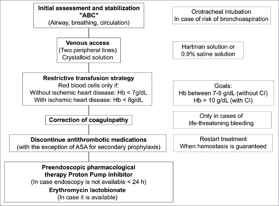
Figure 1. Initial management algorithm for patients with peptic ulcer bleeding. ASA: acetylsalicylic acid; IHD: ischemic heart disease; Hb: hemoglobin.
- – Immediate evaluation of hemodynamic status (airway, breathing, circulation). Consider orotracheal intubation only if there is aspiration risk or airway compromise, and extubation should be performed as soon as clinically safe.
- – Venous access: two peripheral lines; replacement with crystalloid solution, 0.9% sodium chloride saline; consider central access in case of failure.
- – Red blood cell transfusion: in hemodynamically stable patients without ischemic cardiomyopathy, a restrictive transfusion strategy should be considered, that is, transfuse only when hemoglobin is < 7 g/dL; the goal is to maintain a hemoglobin level of 7-9 g/dL post-transfusion. In patients with established cardiovascular disease, the hemoglobin cutoff point to initiate red blood cell transfusion is < 8 g/dL, and the goal is to maintain values > 10 g/dL post-transfusion.
- – Correction of coagulopathy: reversal of anticoagulation should be considered in those patients who present a life-threatening bleeding event (shock, requirement for vasoactive drugs, hemoglobin level decrease > 5 g/dL, transfusion requirement > 5 units of red blood cells). Use of prothrombin concentrate, plasma/platelets, or specific antagonist agents should be considered according to the antithrombotic agent used. This measure should not delay endoscopic evaluation. – Antithrombotic drugs: in patients taking aspirin at cardioprotective doses as primary prevention, it may be suspended and restarted after evaluating its indication. In patients taking it as secondary prevention, it should not be suspended. The rest of antiplatelet and anticoagulant agents should be suspended and their reinitiation performed as soon as possible once endoscopic hemostasis is guaranteed, ideally within the first 5 and 7 days, respectively.
- – Pharmacological therapy prior to endoscopy: intravenous administration of a PPI may be considered before endoscopy if it cannot be performed within the first 24 hours of patient presentation; evidence shows a decrease in high-risk stigmata and need for endoscopic therapies, but does not substitute for endoscopy.
- – Intravenous erythromycin (erythromycin lactobionate if available and not contraindicated) 30-120 minutes before endoscopy, to empty the stomach and improve diagnostic and therapeutic yield.
Endoscopy: optimal timing and classification
Timing of endoscopy
Early endoscopy is considered to be performed within the first 24 hours of admission; in high-risk patients, performing the study within this time decreases the risk of death and need for surgery. The European Society of Gastrointestinal Endoscopy (ESGE) suggests the term “urgent endoscopy” for that performed within the first 12 hours of admission, and “delayed endoscopy” for that performed beyond 24 hours3. In a clinical trial with high-risk patients and suspected non-variceal bleeding, hemodynamically stable, Lau et al.14 reported that, although hemostatic procedures were greater for the endoscopy group in the first 6 hours, bleeding recurrence and 30-day mortality rates were the same in the < 6-hour group and the 6-24-hour group. Currently, international guidelines recommend performing endoscopy within the first 12-24 hours while hemodynamic stabilization is achieved, as “urgent endoscopy” (within the first 12 hours) has not shown mortality benefit in stable patients.2–4,15
Endoscopic classification: Forrest
Since its description by Forrest in 1974, PU classification has remained stable and allows endoscopists to standardize decision-making. Forrest describes three groups: active bleeding (Forrest I), recent bleeding stigmata (Forrest II), and PU without bleeding (Forrest III)16; according to this classification and its subtypes, studies have been conducted determining bleeding recurrence risk for each lesion and the need or not for endoscopic intervention17 (Table 2).
Table 2. Forrest classification
| Type | Description | Bleeding recurrence risk (%) | Risk stratification |
|---|---|---|---|
| Ia | Active arterial bleeding | 80-90 | High risk of bleeding recurrence |
| Ib | Active oozing or spurting bleeding | 40-60 | |
| IIa | Visible vessel without active bleeding | 26 | |
| IIb | Adherent clot | 10-28 | Low risk of bleeding recurrence |
| IIc | Ulcer with hematine-covered base | 13 | |
| III | Ulcer with fibrin-covered base | 4-5 | |
|
Modified from Cappell et al.18. |
|||
Endoscopic treatment
Indications for endoscopic hemostasis
Given the high bleeding recurrence rate without endoscopic treatment, Forrest Ia, Ib, and IIa PUs should be treated.2,3 In a meta-analysis of 19 clinical trials, endoscopic treatment for Forrest Ia, Ib, and IIa significantly reduced bleeding recurrence and surgery rates compared to medical treatment.18,19
For Forrest IIb PUs, studies report that endoscopic treatment reduced bleeding recurrence and surgery rates, but there was no difference in mortality compared to medical treatment; however, other studies report that endoscopic treatment offers no advantages over medical treatment (PPI).2 A clinical trial compared omeprazole with placebo and reported that none of the patients with Forrest IIb PU in the omeprazole group presented bleeding recurrence, required surgery, or had a fatal outcome.20 For this reason, guidelines recommend applying profuse irrigation, and if removal is achieved and Forrest Ia, Ib, or IIa is evidenced, appropriate treatment will be given,3,4 but if the clot remains adherent, no endoscopic therapy may be offered.2
On the other hand, Forrest IIc and III PUs can be managed with medical therapy, as they have low bleeding recurrence risk.2–4
Endoscopic techniques and evidence
Endoscopic therapy can be of four types: injection therapy, mechanical therapy, thermal therapy, and topical therapy.
INJECTION THERAPY
1) Diluted epinephrine
It is used in usual dilution 1:10,000 and is prepared with 1 mL of epinephrine brought to 10 mL with 0.9% saline solution (9 mL). It is suggested to inject in four quadrants adjacent to the bleeding vessel or ulcer edges.1 Hemostasis is achieved by local vasoconstriction, which allows temporary bleeding control, better visualization of the bleeding point, and ultimately greater success when combined with another therapy (thermal or mechanical). The advantages offered are easy application, wide availability, and low cost.1 Due to its temporary nature, it should not be used as monotherapy, as it is associated with a bleeding recurrence of 12-30%.1–4,19
2) Absolute alcohol injection
This method is based on the principle of tissue dehydration and fixation with absolute alcohol, which produces vasoconstriction and vascular wall necrosis, favoring thrombogenesis and hemostasis. The recommendation is to inject aliquots of 0.1-0.2 mL of absolute alcohol adjacent to the bleeding site; a change in blood vessel color to brown indicates appropriate hemostasis. To avoid perforation, it is recommended not to exceed 1.5 mL. It is not recommended as monotherapy and its use should be avoided in PU with active bleeding.2,4
MECHANICAL THERAPY
1) Through-the-scope (TTS) mechanical clips
They are especially indicated on stable and accessible edges. Firm and precise clip placement is important to avoid bleeding recurrence. They offer therapeutic success rate of 85-100%. They have the advantages of wide availability, easy use, and low cost. Unlike thermal therapy, they avoid tissue damage and thus late perforation risk; however, their therapeutic success decreases in PU > 2 cm and those located on the lesser curvature or posterior duodenal wall.1
2) Over-the-scope clips (OTSC)
It is a clipping system that is mounted on the endoscope (similar to endoscopic band ligation). It was initially designed for tissue closure (fistulas, perforations), but its usefulness has been demonstrated as endoscopic therapy in cases of bleeding recurrence, even superior to conventional therapy, and recently its usefulness in PU has been confirmed as first-line therapy in patients at high risk of bleeding recurrence (RS ≥ 7).1,4,11 It is recommended to use atraumatic OTSC clips, as they are associated with lower bleeding recurrence rates (7.6% vs. 34.9%) and fewer transfusion requirements (2.5 vs. 5.1 units) compared to traumatic ones.11 The disadvantages associated with this therapy are cost and availability. ESGE recommends its use as first line in ulcers > 2 cm with visible vessel > 2 mm, at sites with fibrosis, and on broad bases,3 while the American College of Gastroenterology still suggests them as rescue therapy in cases of bleeding recurrence.11
THERMAL THERAPY
1) Contact therapy
Various devices exist that can apply heat to cauterize the bleeding site. According to their operating mechanism, they can be divided into two types:
– Bipolar current: the tip of a hot probe is placed on the bleeding site and slight pressure is applied to the tissue, constantly for 8-10 seconds, to achieve hemostasis.1
– Monopolar current: hemostatic forceps (hot biopsy forceps, coagrasper) with low-voltage coagulation can be an effective option for PU treatment, especially when it is difficult to locate or has a rigid or fibrotic base.1
2) Non-contact therapy
Argon plasma coagulation is the preferred option when hemostasis is required superficially or diffusely. The operator must have precise endoscope control to maintain a distance of 1-2 mm between the applicator tip and tissue, thereby avoiding submucosal emphysema or late perforation.1,4
For heat application with any device, the bleeding site must be identified, and thermal therapy application “blindly” or with limited visibility should be avoided at all costs.
TOPICAL THERAPY
This therapy modality is relatively new and is generally used as adjuvant therapy, as a last alternative when other hemostatic methods fail, or as bridge therapy when conventional treatments are not possible.1,3,21 It has recently been proposed as a first-line option, but there is still no conclusive evidence favoring its use as first-choice therapy.
International guidelines agree that topical hemostatic powder therapy (specifically Hemospray®) is useful as a temporary rescue measure when conventional endoscopic therapy fails in non-variceal upper gastrointestinal bleeding, but should not be used as sole treatment. Its use is recommended as primary therapy in tumor or massive bleeding when other treatments are not feasible.21
- 1. Thrombin/fibrin-derived products
- Generally sprayed over the bleeding site to favor clot formation. They are used for diffuse or oozing bleeding.
- 2. Hemostatic powders
- – TC-325 (Hemospray®): it is a mineral hemostatic powder (sodium bentonite) that upon contact with the bleeding site absorbs water and forms a mechanical barrier by aggregation and adhesion, promoting clot stability, controlling bleeding. Clinical trials have demonstrated non-inferiority compared to conventional therapy, with high immediate hemostasis rates and low bleeding recurrence risk. The applicator is easy to use and does not require much precision; however, it requires active bleeding to be effective, and once applied, it limits endoscopic vision.1
- – EndoClot®: it is a hemostatic polysaccharide derived from starch. It has efficacy similar to Hemospray®, with no difference in bleeding recurrence rates.1 Clinical trials have demonstrated non-inferiority compared to conventional treatment in terms of bleeding recurrence rates and hemostatic success.21
- – Nexpowder®: it is an inert blue biocompatible polymer composed of aldehyde dextran and succinic acid-modified ε-poly(l-lysine), which forms an adhesive hydrogel and functions as a mechanical barrier. It does not require active bleeding for adherence.1,21
- – PuraStat®: it is a self-assembling synthetic peptide that forms a transparent hydrogel barrier on contact with blood or moisture, sealing the bleeding site and promoting hemostasis.1 A meta-analysis has reported hemostatic success of 93.1%, with a bleeding recurrence rate of 8.9%.21 In a multicenter prospective study with 111 patients, efficacy of 94% was found when used as first-line therapy, and 75% as rescue therapy, with a bleeding recurrence rate of 16% at 30 days.11
Combined therapy: which method to choose?
Although characteristics and risk factors inherent to each patient must be evaluated individually, therapeutic success is estimated to be > 90% for all methods and bleeding recurrence is approximately 2-10%, except for epinephrine injection.3,4
In several studies, it has been observed that the combination of two endoscopic therapies (injection plus thermal therapy or injection plus mechanical therapy) is more effective in reducing bleeding recurrence than monotherapy.4,11,19
In a clinical trial, hemostatic treatment with hot biopsy forceps in “soft coagulation” mode had a higher initial hemostasis rate, lower bleeding recurrence rate, and shorter hospital stay compared to hemoclips after epinephrine injection for active bleeding.22 A clinical trial found no difference between argon plasma coagulation and hemoclips.23
Endoscopic treatment will depend not only on PU characteristics but also on patient risk factors (anticoagulant or antithrombotic use, comorbidity, etc.), endoscopist skills, and resources available in the endoscopy unit.4 Figure 2 shows a therapeutic algorithm according to current evidence.
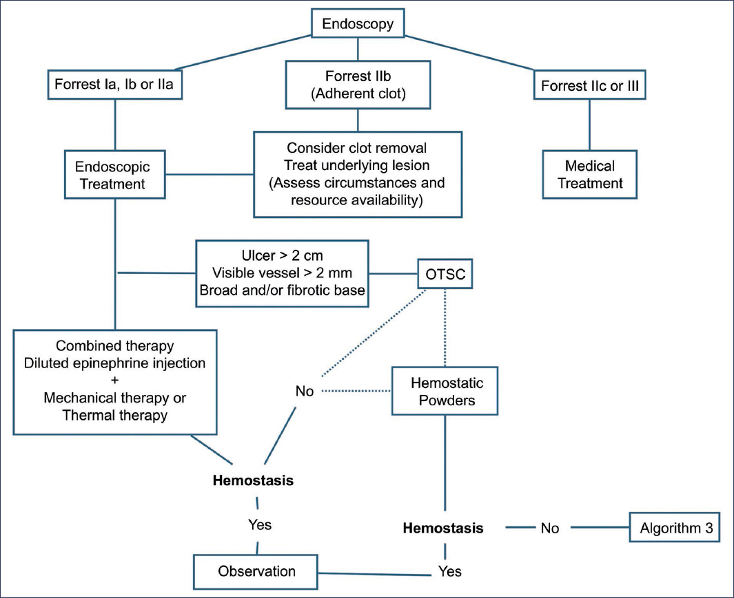
Figure 2. Therapeutic algorithm for patients with peptic ulcer bleeding. OTSC: over-the-scope clips.
Pharmacological therapy after endoscopy
Proton pump inhibitors
In patients with high-risk lesions who were treated endoscopically, an intravenous PPI will be administered at a dose of 80 mg initial bolus followed by continuous infusion of 8 mg/h for 72 hours. Several meta-analyses have tested high-dose intermittent PPI use (80 mg daily, intravenously or orally) and results are similar in terms of bleeding recurrence, need for radiological or surgical treatment, and mortality with continuous dosing of 8 mg/h.3 Subsequently, double-dose PPI may be continued intravenously or orally for 2 weeks.
H. pylori eradication
H. pylori eradication is indicated in patients with active or previous peptic ulcer. This measure reduces ulcer recurrence and its associated complications.
Although the rapid urease test is considered an accurate, easy-to-use, and rapid diagnostic method to detect active H. pylori infection, its usefulness in the context of active bleeding is limited due to the high probability of obtaining false-negative results.24
Management of NSAIDs, antiplatelet agents, and anticoagulants
After a gastrointestinal bleeding event, NSAIDs should be temporarily suspended and their reinitiation requires evaluation of their indication and risk-benefit of their use. If essential, use of selective cyclooxygenase-2 inhibitors or concomitant PPI use may be considered.10
It is important to note that in patients with gastrointestinal bleeding, aspirin is not recommended to be suspended when used at cardioprophylactic doses for secondary prevention. If for any reason it was suspended, it should be restarted as soon as possible.25
After a gastrointestinal bleeding event, antithrombotic therapy reinitiation is recommended, when indicated, in patients with acute coronary syndromes or atrial fibrillation, or after percutaneous intervention. However, the timing of reinitiation is critical, as premature reintroduction may generate bleeding recurrence, while late reintroduction may increase thromboembolic event risk. Antithrombotic therapy decreases unfavorable outcomes secondary to underlying etiology.26 The timing of treatment reinitiation after gastrointestinal bleeding requires an individualized approach in each patient, and in some cases coordination with other specialists in cardiology, neurology, or hematology will be required. In general, antiplatelet agents and anticoagulants should be restarted as soon as clinically safe during the first days after suspension.27,28
Management of bleeding recurrence
If the patient presents signs of bleeding recurrence, such as hemodynamic instability, persistent hematemesis or profuse melena, or significant hemoglobin decrease (> 2 g/dL), bleeding recurrence should be considered and endoscopic treatment offered as first option.2 The therapeutic algorithm when there is evidence of bleeding recurrence is shown in figure 3.
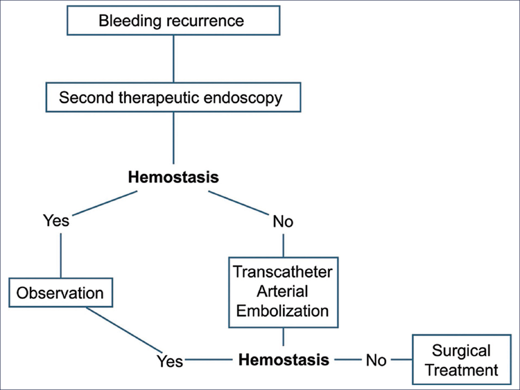
Figure 3. Therapeutic algorithm for patients with recurrent peptic ulcer bleeding.
Second therapeutic endoscopy
A second endoscopy (second look) is not recommended as part of PU treatment3; however, if there are clinical signs of bleeding recurrence, such as hemodynamic instability, a second endoscopy for therapeutic purposes is justified. Early second endoscopy (24 hours) with additional therapy or technique change (e.g., OTSC) is effective in a high percentage of cases.2
Ulcers > 2 cm and presence of shock are predictive factors for endoscopic retreatment failure.4
Radiological therapy
In patients with persistent or refractory bleeding to endoscopic treatment, transcatheter arterial embolization is recommended,2–4 which achieves technical hemostasis of 90-100%, but clinical hemostasis of 50-83%, and not always permanently. Coagulopathies, massive blood transfusion, and long-duration procedures are risk factors for bleeding recurrence after transcatheter arterial embolization.4 Studies have demonstrated higher bleeding recurrence rates compared to surgery, but fewer complications.29 The choice depends on anatomy, stability, and local experience.
Surgery
Surgical treatment is indicated when endoscopic and endovascular methods fail, when transcatheter arterial embolization is not available, and in massive uncontrollable bleeding.3,4
Secondary prevention and follow-up
Eradicating H. pylori restores normal acid secretion, allowing ulcer healing and preventing relapse. Among patients not treated for H. pylori, recurrence rates were 52% for gastric ulcer and 64% for duodenal ulcer. Among patients with successful H. pylori eradication, annual ulcer recurrence is approximately 0% to 2.3% for gastric ulcer, and 0% to 1.6% for duodenal ulcer, in a follow-up period of up to 9.8 years6. The Fifth Mexican Consensus on the diagnosis and treatment of H. pylori infection recommends demonstrating its eradication 4 weeks after treatment, through a non-invasive method.24
Suspending NSAID use in people who develop an ulcer while using these drugs allows healing of the gastric mucosal barrier, restoring normal protective mechanisms. In patients with gastrointestinal risk factors, avoiding NSAIDs is recommended, but when essential, H. pylori eradication, use of selective cyclooxygenase-2 inhibitors, or concomitant PPI use is suggested.6,10,24
In patients with high thrombotic risk, interruption of antithrombotic treatment after a UGIB event increases thrombotic complication risk and mortality. Resumption of antiplatelet agents and anticoagulants should be individualized, balancing bleeding recurrence risk against thrombosis risk. Resumption of antithrombotic treatment has become a key intervention, critical for patient outcomes in UGIB, and when pertinent, patients on antithrombotic treatment should have an antithrombotic plan after endoscopy for UGIB.
Perspectives and emerging technologies
Red dichromatic imaging is an optical filter designed by Olympus, available with EVIS-X1 processors, which improves contrast between concentrated and diluted blood, allowing better identification of the bleeding site for endoscopic treatment. There are still no clinical trials reporting its efficacy in terms of greater bleeding control.11
Endoscopic suturing has emerged as another alternative to achieve hemostasis in large PUs, particularly in those at high perforation risk or refractory to conventional treatment. In a prospective study conducted in China, approximation of ulcer edges with an endoloop was used as first-line treatment in ulcers > 1 cm, with hemostatic success of 89% and clinical success in PU refractory to conventional therapy of 81.2%. On the other hand, various case reports exist using suturing devices, such as Apollo OverStitch®; however, more studies are still needed to recommend their use.11
Conclusions
In a patient with UGIB, priorities are rapid evaluation, hemodynamic stabilization, risk stratification, and a restrictive transfusion strategy. Early endoscopy (< 24 hours) is indicated for most patients, while urgent endoscopy (< 12 hours) is reserved for unstable patients. Combined endoscopic therapy (diluted epinephrine injection plus thermal or mechanical method) should be considered for high-risk lesions (Forrest Ia, Ib, and IIa). OTSC use can be considered in refractory ulcers or with large visible vessel; growing evidence is emerging for its use as an initial measure, but is still in consolidation. In cases of bleeding recurrence, a second endoscopy is suggested as first line; if it fails, consider radiological therapy or surgery according to availability.
Funding
The authors declare having received no funding for this study.
Conflicts of interest
The authors declare having no conflicts of interest.
Ethical considerations
Protection of people and animals. The authors declare that no experiments were conducted on human beings or animals for this research.
Confidentiality, informed consent, and ethical approval. The study does not involve personal data, clinical histories, or human biological samples, so it does not require ethical approval. SAGER guidelines do not apply.
Declaration on the use of artificial intelligence. The authors declare that no type of generative artificial intelligence was used for the writing or creation of content of this manuscript.
