
Introduction
Gastritis is defined by the histological presence of inflammation in the gastric mucosa produced by infectious agents or autoimmune causes, as well as the patient’s response to these lesions. It should be mentioned that epithelial injury in the mucosa (with or without regeneration) without inflammation (host response) is termed “gastropathy”; however, these two phenomena may coexist and clinically usually present similarly. The first description of gastritis emerged in the 19th century as a result of autopsy analysis, and in 1947 it was categorized as acute and chronic.1 Although there were different stratification systems, such as the Schindler classification, with the advent of evidence of Helicobacter pylori as a causative agent of gastritis in 1983, progress was made toward an integrated classification of endoscopic, histological, and etiological findings: the Sydney classification.2
Gastritis can be classified according to temporality, histological characteristics, and etiology, but there is no universally accepted classification, and for the purposes of clinical practice, it is essential to understand the pathophysiological mechanisms in order to provide a clinical management plan. This review will address gastritis from an etiological, clinical, and general therapeutic point of view.
Methodology
A narrative review focused on evidence published in international guidelines and consensus statements from the last 5 years was conducted, focusing on definition, etiology, and pharmacological treatment strategies.
Etiology and classification of histopathological findings
The etiological approach to gastritis classifies it into three main groups: acute, chronic, and special. However, with the integration of clinical context, endoscopic findings, and etiology, the classifications shown in tables 1 and 2 can be considered. Furthermore, histological classification is sometimes challenging for the clinician to understand in decision-making. Table 3 shows the classification derived from the revision of the Sydney system in 19943,4 according to the main findings obtained in histopathology reports, which may vary according to the etiology and temporality of gastritis. Of particular interest is the presence of atrophic changes with disappearance or substitution of gland types toward gastric dysplasia; thus, the report in these cases should employ the OLGA (Operative Link Gastritis Assessment) system,5,6 shown in table 4; stages III and IV are particularly important, as they carry considerable risk for the development of gastric cancer and require the implementation of an endoscopic surveillance program.7
Table 1. Etiology of gastritis
| Induced by Helicobacter pylori | Special types of gastritis Allergic Secondary to bile reflux Lymphocytic Eosinophilic Ménétrier disease |
| Induced by medications Nonsteroidal anti-inflammatory drugs Iron supplements Bisphosphonates |
|
| Autoimmune | Secondary to external causes Alcohol Radiation Chemical |
| Stress-mediated Sepsis Hypovolemia Drug abuse (cocaine) |
|
| InfectiousPyogenic (phlegmon) | Crohn disease |
| Sarcoidosis | |
| Bacterial Enterococci Mycobacteria (tuberculosis and non-tuberculosis) Syphilis | Vasculitis |
| Viral Cytomegalovirus Enterovirus | |
| Fungal Mucormycosis Candidiasis Histoplasmosis | |
| Parasitic Anisakiasis Cryptosporidium Strongyloides stercoralis | |
Table 2. Classification of gastritis and examples of etiology
| Group | Etiology |
|---|---|
| Acute | Drugs, stress-induced, uremia, ischemia, shock, corrosive agents, radiation, irritative (foods), sepsis, trauma, bacterial and viral infections, alcohol use, burns, bile/alkaline reflux, major surgery, organ failure, portal hypertension, congestive heart failure, increased intracranial pressure |
| Reactive gastropathy (chemical) | Endotoxic (bile/alkaline reflux, uremia)Exotoxic (nonsteroidal anti-inflammatory drugs, alcohol)Stress-induced |
| Chronic | Helicobacter pylori (and Helicobacter heilmannii)AutoimmuneChronic H. pylori-negative gastritis |
| Special | LymphocytiCollagenouscEosinophilicRadiationGraft-versus-host diseaseBacterial (syphilis, tuberculosis)Viral (cytomegalovirus, herpes simplex virus)Fungal (Candida, Aspergillus, Mucormycosis, Coccidioides, Histoplasma, Cryptococcus, Pneumocystis)Parasitic (Anisakis, Cryptosporidium, Ascaris lumbricoides, Giardia, Toxoplasma, Schistosoma) |
| Granulomatous | IdiopathicCrohn diseaseSarcoidosisBarium granulomas |
| Hypertrophic | Ménétrier diseaseZollinger-Ellison syndromeHypertrophicHypersecretory (protein-losing enteropathy) |
| Gastric vasculopathies | IschemicGastric antral vascular ectasia (watermelon stomach)Portal hypertensive gastropathy (congestive gastropathy)VaricesAngiodysplasiaDieulafoy lesionTelangiectasias associated with hemodialysis |
| Gastric involvement in systemic diseases | Inflammatory bowel diseaseAmyloidosisDiabetes>MastocytosisSjögren syndromeHypercalcemiaSiderosis |
|
Translated from Pennelli et al.12. |
|
Table 3. Histopathological findings in gastric biopsy pathology reports
| Criterion | Mechanism/relevance | Normal finding | Measurement |
|---|---|---|---|
| Mononuclear leukocyte infiltrate | Intensity of gastritis | 2 to 5 lymphocytes, plasma cells, or macrophages per high-power field 2 or 3 cells between foveolae Lymphocytes: < 25/100 epithelial cells |
Mild (+) Moderate (++) Severe (+++) Lymphocytes: 25/100 |
| Neutrophil infiltrate | Gastritis activity | 2 to 5 neutrophils per high-power field 2 or 3 neutrophils |
Location: Lamina propria (+) Epithelium (++) Glandular lumen (+++) |
| Helicobacter pylori density | Presence (and quantity) of H. pylori | Presence or absence | Mild Moderate Severe |
| Foveolar hyperplasia | Expansion of gastric glands and presence of dysplasia | Description of regenerative changes in glandular epithelium | Difference from dysplastic lesions: Indefinite for dysplasia Atypical foveolar hyperproliferation |
| Gastric atrophy | Loss of gastric glands (mucosecreting or oxyntic) | Atrophy should not be present Two subtypes: Non-metaplastic: Gland disappearance with sclerosis of lamina propria Metaplastic: Gland replacement: intestinal metaplasia vs. pseudopyloric metaplasia (oxyntic glands) |
1 = 1-30% specimens 2 = 31-60% specimens 3 = > 60% specimens |
| Endocrine cell hyperplasia | Enterochromaffin cell hyperplasia | Hyperplasia and cell alignment should not exist | Precursor of gastric neuroendocrine tumors Alignment of 5 enterochromaffin cells in lamina propria |
| Lamina propria fibrosis and muscularis mucosae hypertrophy | Scarring following ulcerative or inflammatory processes | Relevant to distinguish from mucosal atrophy | Indicates scarring process |
| Oxyntic epithelium hypertrophy | Hypertrophic changes in oxyntic epithelium | Usually related to use of proton pump inhibitors and in atrophic gastritis | Report with uncertain clinical significance |
Table 4. OLGA reporting system
| Global atrophy score | Atrophy score in oxyntic tissue (body and fundus) | |||
|---|---|---|---|---|
| 0: no atrophy | 1 | 2 | 3 | |
| 1: 1-30% specimens | ||||
| 2: 31-60% specimens | ||||
| 3: > 60% specimens | ||||
| 0 | ||||
| Atrophy score in mucosecretingtissue (antrum and angular incisure) | 0 | No stage | Stage I | Stage II |
| 1 | Stage I | Stage I | Stage II | |
| 2 | Stage II | Stage II | Stage III | |
| 3 | Stage III | Stage III | Stage IV | |
|
Translated from Pennelli et al.19. |
||||
Diagnosis
The clinical presentation of gastritis is usually conditioned by the clinical and etiological context in which a given patient is evaluated, although it is usually nonspecific and usually not determinative of etiology. Typical symptoms usually include epigastric pain or burning, fullness, early satiety, nausea, and vomiting, although occasionally it may be found incidentally during endoscopic evaluation. Thus, the differential diagnosis includes conditions such as functional dyspepsia, peptic ulcer disease, cholecystitis, pancreatitis, myocardial infarction, and celiac disease, among other upper gastrointestinal tract pathologies. In this way, diagnostic management and approach will be largely conditioned by the clinical context and, above all, by the presence of alarm signs or symptoms, some of which are age over 55 years, unexplained weight loss, presence of anemia, melena, persistent vomiting, and family history of gastric cancer.8,9
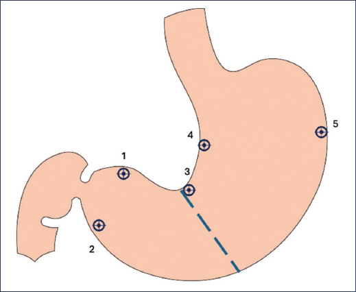
Upper endoscopy is a fundamental test in the diagnosis of gastritis, since without it, it is not possible to obtain the histopathological specimens that allow differential diagnosis of gastritis. Although it is essential to provide a description and photographic documentation of all segments and findings during the study (such as presence of focal lesions, fold thickening, polyps, vasculature, masses, or erosions) (Fig. 1), these findings usually have limited value because there is great interobserver variability.10 Furthermore, it has been shown that the sensitivity of endoscopic findings in relation to the histopathological diagnosis of gastritis or H. pylori infection is approximately 57%.11 On the other hand, although there may be local variability in the number of biopsies (according to available resources and technology, as well as clinical context), it is generally recommended to follow the standard Sydney protocol for performing biopsies, according to which one or two biopsies are taken from five gastric anatomical zones: greater curvature of the antrum, lesser curvature of the antrum, angular incisure, greater curvature of the body, and lesser curvature of the body (Fig. 2). This protocol usually allows optimization of etiological diagnosis.
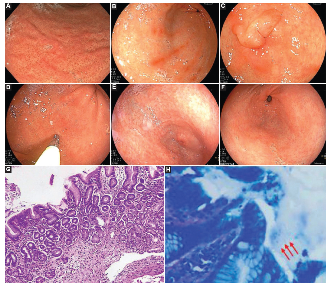
Figure 1. Common endoscopic and histopathological findings in patients with gastritis (images courtesy of Dr. Genaro Vázquez-Elizondo). A: mucosa with edematous appearance and erythema. B: linear erosions in the antrum and body. C: prepyloric erythema. D: diffuse erythema in the body. E: erythema and flattened changes in the antrum and body. F: minimal mucosal changes. G: mononuclear infiltrate typical of gastritis (hematoxylin and eosin stain). H: bacillary structures compatible with Helicobacter pylori (Diff-Quick stain). All biopsies from these patients showed the presence of this microorganism (red arrows).
Treatment
Regarding the treatment of erosive gastritis, we must recognize that, although the etiology may be multifactorial – as previously explained – current international and national clinical practice guidelines direct treatment toward specific causes, which typically include eradication of H. pylori, reducing or preventing toxicity from drugs such as nonsteroidal anti-inflammatory drugs, and the use of acid-blocking agents such as H2 receptor blockers, proton pump inhibitors, or potassium-competitive acid blockers, according to the specific clinical context.8,12–16 Additionally, for purely symptomatic control, it is recommended to combine these pharmacological options with general measures, which include suspension of precipitating dietary elements and reviewing the appropriateness and indication of some drugs (such as acetylsalicylic acid and iron, among others).
One of the most frequent considerations when treating patients with gastritis is the eradication of H. pylori, which is recommended in those with evidence of atrophic gastritis or intestinal metaplasia, since it has been shown to stop the progression of neoplastic changes.17 In patients with dyspeptic symptoms, eradication is a first-line treatment, as it improves the magnitude of symptoms.8,12 Recently, the Mexican Association of Gastroenterology published the Fifth Mexican Consensus on the Diagnosis and Treatment of Helicobacter pylori Infection, in which the guidelines for this therapeutic option can be consulted.18 Regarding other pharmacological alternatives, there is evidence for the use of mucoprotective agents, acid blockers, and cytoprotective agents. Table 5 summarizes the options available in Mexico.
Table 5. Pharmacological alternatives for the management of gastritis
| Drug | Mechanism of action | Type of available studies | Doses evaluated | Clinical outcomes | Comments |
|---|---|---|---|---|---|
| Rebamipide | Prostaglandin stimulation, reduction of oxidative stress and modulation of inflammatory cytokines | 9 RCTs; 1 prospective study, 1 comparative study with active agent | 300 mg/day divided doses for 2 and up to 26 weeks (8 weeks standard) | Improvement in symptom score, endoscopic lesions, and histological inflammation indices | Studied in erosive and chronic gastritis Effect superior to sucralfate and similar to famotidine |
| Sucralfate | Cytoprotective agent that forms a barrier over damaged mucosa promoting healing through stimulation of prostaglandins and mucus | 3 RCTs, 2 placebo-controlled | 4 to 6 g/day divided doses (6 and up to 12 weeks) | Histological improvement in chronic non-erosive bile-mediated gastritis post- cholecystectomy), but no effect on dyspeptic symptoms | Borderline effect compared to ranitidine Effect inferior to rebamipide |
| Famotidine | H2 receptor antagonist in parietal cells | 3 RCTs comparative with active compound; 1 escalating dose comparative study | 5 to 20 mg/day in different doses for 2 weeks | Non-inferior effect to esomeprazole and rebamipide | Used for short periods and not evaluated beyond 2 weeks |
| Ranitidine | H2 receptor antagonist in parietal cells | 4 comparative RCTs with active compound | 300 mg divided doses for 8 weeks; up to 6 months in maintenance | Consistent effect vs. placebo, but inferior to proton pump inhibitors in erosions and histology Superior to sucralfate in symptoms and histology | Effect superior to sucralfate, but less than other acid-suppressing agents |
| Omeprazole | First-generation proton pump inhibitor | 2 comparative and multicenter RCTs in patients using nonsteroidal anti-inflammatory drugs and ulcers or > 10 erosions | 20 to 40 mg/day for 4 to 8 weeks; up to 6 months maintenance | Effect superior to misoprostol and ranitidine in erosions at 4 and 8 weeks | Evidence in patients with nonsteroidal anti-inflammatory drug toxicity |
| Esomeprazole | Second- generation proton pump inhibitor | Comparative RCT with famotidine | 10 mg daily for 2 weeks | Effect similar to 20 mg ranitidine | Reduced doses in patients with erosive gastritis |
| Rabeprazole | Second-generation proton pump inhibitor | 3 comparative RCTs in patients with endoscopic and histological gastritis Evaluated in reactive (bile) gastritis | 20 mg/day for 4 to 12 weeks | Effect similar to sucralfate in biliary gastritis Effective in chronic non-ulcerative gastritis | Limited evidence and not superior to sucralfate |
| Tegoprazan | Potassium- competitive acid blocker | 2 phase III RCTs compared to placebo | 10 to 20 mg/day divided doses for 2 weeks | Consistent effect and superior to placebo for short periods | Still no comparative studies against active compound |
|
RCT: randomized controlled trial. Developed from Kim et al.2. |
|||||
Conclusions
Erosive gastritis is a clinical-endoscopic condition with multifactorial etiology. The optimal approach integrates: identifying and treating the cause (especially H. pylori and exposure to nonsteroidal anti-inflammatory drugs) and suppressing acid and protecting the mucosa when indicated. Acute and chronic management of this condition, including follow-up studies, will largely depend on the specific clinical context.
Funding
The author declares not having received funding for this study.
Conflicts of interest
The author declares having no conflicts of interest.
Ethical considerations
Protection of human and animal subjects. The author declares that no experiments were performed on humans or animals for this research.
Confidentiality, informed consent, and ethical approval. The study does not involve personal data, medical records, or human biological samples, so ethical approval is not required. SAGER guidelines do not apply.
Declaration on the use of artificial intelligence. The author declares that no type of generative artificial intelligence was used for the writing or creation of content of this manuscript.
