
Introduction
Peptic ulcer disease (PUD) represents one of the classic models in gastroenterology for studying the interaction between luminal aggressive factors and the protective mechanisms of the gastrointestinal mucosa.1,2 Throughout much of the 20th century, its pathophysiology was primarily explained based on the concept of acid hypersecretion, which led to considering hydrochloric acid as the main determinant of mucosal damage and directing therapeutic strategies almost exclusively toward its suppression.3 This approach enabled significant advances in the clinical management of the disease, but proved insufficient to explain why not all individuals exposed to similar levels of acid secretion develop mucosal lesions.4
The recognition of the etiopathogenic role of Helicobacter pylori and the damage induced by nonsteroidal anti-inflammatory drugs (NSAIDs) marked a turning point in the understanding of PUD.5–7 These findings demonstrated that mucosal aggression depends not only on acid but also on a complex interaction of chemical, inflammatory, immune, and structural factors that compromise the integrity of the gastroduodenal barrier.8,9 In parallel, knowledge of mucosal defense mechanisms has deepened, including the mucus and bicarbonate layer, prostaglandins, adequate mucosal blood flow, epithelial restitution capacity, and regulation of the local immune response.10–12
Currently, PUD is defined as the result of a dynamic imbalance between aggressive factors and mucosal defense systems, modulated by individual host susceptibility.2,13,14 This multidimensional pathophysiological model allows us to explain clinical variability, the persistence of damage despite acid suppression, and disease recurrence in certain patients.14,15 Understanding these mechanisms is fundamental not only for a more precise interpretation of pathogenesis but also for the development of more effective therapeutic and preventive strategies.
The objective of this article is to provide an updated review of the pathophysiology of PUD, integrating the mechanisms of aggression and defense of the gastroduodenal mucosa and analyzing their clinical relevance in light of current scientific evidence.
Current pathophysiology of peptic ulcer disease
Mucosal balance and gastroduodenal homeostasis
The gastroduodenal mucosa is constantly exposed to a highly hostile environment, characterized by the presence of hydrochloric acid, pepsin, and other potentially harmful factors. Despite this, under physiological conditions, the structural and functional integrity of the mucosa is maintained thanks to a complex and dynamic system of defensive mechanisms that preserve local homeostasis.16,17 This concept of mucosal balance constitutes the basis of the modern understanding of PUD and reshapes the view focused exclusively on acid secretion (Fig. 1).
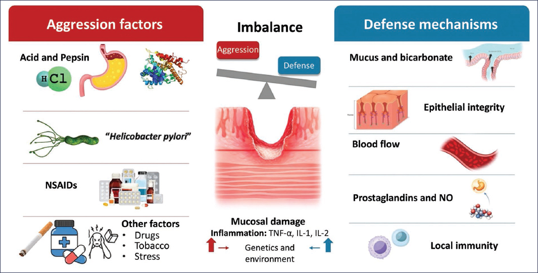
Figure 1. Pathophysiological model of the balance between aggression factors and gastric mucosal defense mechanisms. The integrity of the gastric mucosa depends on the dynamic balance between aggressive factors – hydrochloric acid and pepsin, Helicobacter pylori, nonsteroidal anti-inflammatory drugs (NSAIDs), and other factors such as tobacco, stress, and drugs – and mucosal defense mechanisms, which include mucus and bicarbonate secretion, epithelial integrity, adequate blood flow, production of prostaglandins and nitric oxide, as well as local immunity. Imbalance in favor of aggression conditions mucosal damage and inflammation, mediated by proinflammatory cytokines (TNF-α, IL-1, IL-2), modulated by genetic and environmental factors, favoring the development of ulcerative lesions.
Gastroduodenal homeostasis depends on the coordinated interaction of aggression and defense factors, modulated by the adaptive capacity of the epithelium and the host’s inflammatory response. In this context, gastric acid should not be considered a pathological agent per se, but rather an essential physiological component for digestion and defense against pathogens, whose harmful potential is only manifested when mucosal protection mechanisms are overwhelmed or compromised.18 This notion explains why individuals with normal levels of acid secretion can develop PUD, while others with hypersecretion remain free of mucosal damage.
The gastric and duodenal epithelium functions as a highly specialized barrier, capable of self-regulating in response to noxious stimuli through pre-epithelial, epithelial, and sub-epithelial defense mechanisms. The mucus and bicarbonate layer constitutes the first line of protection, generating a microenvironment with an almost neutral pH at the epithelial surface, even in the presence of a highly acidic lumen.19 This pH gradient is fundamental for limiting pepsin activation and reducing direct damage to epithelial cells.
In the epithelium, the integrity of tight junctions and the capacity for cell restitution allow rapid repair of acid-induced microlesions or by other aggressive agents. Epithelial restitution, a process independent of cell proliferation, occurs within minutes to hours and depends on local signals mediated by growth factors, prostaglandins, and adequate mucosal blood supply.20,21 Disruption of these mechanisms, as occurs with NSAID use or in chronic inflammation, favors progression of damage toward established ulcerative lesions.
Mucosal blood flow plays a central role in maintaining gastroduodenal homeostasis by ensuring oxygen and nutrient supply, facilitating removal of retrogradely diffused protons, and allowing transport of cytoprotective mediators. Blood flow regulation depends largely on prostaglandins and nitric oxide, whose decrease is associated with greater mucosal vulnerability to aggressive factors.22 Mucosal ischemia, even transient, can alter this delicate balance and potentiate acid-induced damage.
From a comprehensive perspective, mucosal balance does not represent a static state, but rather a dynamic process of continuous adaptation to luminal, microbial, and inflammatory stimuli. Some individual host factors, such as genetics, age, the presence of comorbidity, and the immune response, significantly influence the mucosa’s capacity to maintain this homeostasis.23,24 Thus, PUD arises when the sum of aggressions exceeds local defensive capacity, either through increased harmful factors, deterioration of protection mechanisms, or a combination of both.
This pathophysiological model of mucosal balance and imbalance provides a solid conceptual framework for understanding the clinical heterogeneity of PUD and constitutes the basis for analyzing, in the following sections, the specific role of the different aggression factors and defense mechanisms involved in its development and progression.
Aggression factors
The development of PUD is determined by the combined action of various aggressive factors capable of altering the integrity of the gastroduodenal mucosa when they exceed local defense mechanisms. These factors include luminal components, infectious agents, and drugs, whose pathophysiological relevance varies according to individual host susceptibility and clinical context.25
HYDROCHLORIC ACID AND PEPSIN
Hydrochloric acid constitutes an essential physiological element for protein digestion and defense against microorganisms; however, its harmful potential is manifested when the mucosal barrier is compromised. Acid-induced damage occurs primarily through retrograde diffusion of protons toward the epithelium, which leads to alterations in intracellular pH, mitochondrial dysfunction, and activation of cell injury pathways.26 Pepsin, whose activity depends on an acidic medium, amplifies this damage by degrading structural proteins of the mucosa when surface pH decreases in a sustained manner.27
Contrary to the classic model, numerous studies have shown that most patients with PUD have acid secretion levels within normal ranges, which reinforces the idea that acid acts as a necessary, but not sufficient, factor in pathogenesis.28 This finding is particularly evident in ulcers associated with H. pylori or NSAID use, where mucosal damage occurs even without significant hypersecretion.
HELICOBACTER PYLORI
H. pylori infection represents one of the most important aggressive factors in the pathophysiology of PUD. This gram-negative microorganism colonizes the gastric mucosa and triggers a chronic inflammatory response characterized by infiltration of immune cells, release of proinflammatory cytokines, and alteration of epithelial defense mechanisms.29 Infection persistence leads to a state of chronic inflammation that increases mucosal vulnerability to acid and pepsin.
The effects of H. pylori on acid secretion are complex and depend on the pattern of gastritis induced. While antral gastritis is associated with increased acid secretion and higher risk of duodenal ulcer, corporal gastritis can lead to hypochlorhydria and predispose to mucosal damage through inflammatory mechanisms and epithelial barrier alteration.30,31 Additionally, bacterial virulence factors, such as CagA and VacA, directly contribute to tight junction disruption and cell apoptosis, exacerbating mucosal damage.32
NONSTEROIDAL ANTI-INFLAMMATORY DRUGS
NSAID use constitutes another main cause of PUD. Their harmful effect is due to both systemic and topical mechanisms. Cyclooxygenase inhibition, particularly cyclooxygenase-1, reduces the synthesis of gastroprotective prostaglandins, which translates into decreased mucus and bicarbonate secretion, reduced mucosal blood flow, and deterioration of epithelial restitution.33
Additionally, NSAIDs can exert a direct topical effect on the epithelium by altering cell membrane permeability and facilitating proton diffusion toward the interior of epithelial cells, which potentiates acid-induced damage.34 The coexistence of H. pylori infection and NSAID use synergistically increases the risk of developing ulcers and complications, emphasizing the multifactorial nature of mucosal aggression.35
OTHER AGGRESSIVE FACTORS
Other elements, such as duodenogastric reflux, oxidative stress, mucosal ischemia, and certain systemic conditions, can contribute to gastroduodenal damage by altering the balance between aggression and defense. Although their role is usually secondary, these factors acquire relevance in patients with comorbidity or in contexts of prolonged physiological stress, in which the mucosa’s adaptive capacity is diminished.36
Collectively, aggression factors act interdependently and their impact on the mucosa depends not only on their intensity but also on the integrity of local defense mechanisms. This dynamic interaction explains the clinical variability of PUD and prepares the ground for analyzing, in the next section, the defense systems that counteract these aggressions.
Gastroduodenal mucosal defense mechanisms
The integrity of the gastroduodenal mucosa depends on a highly specialized system of defensive mechanisms that act in a coordinated manner to counteract the constant action of aggressive factors. These mechanisms, in addition to limiting acid- and pepsin-induced damage, allow rapid epithelial repair, preserving barrier function and local homeostasis.37
PRE-EPITHELIAL BARRIER: MUCUS AND BICARBONATE
The mucus layer covering the gastric surface constitutes the first line of defense against luminal content. This viscoelastic gel, secreted by superficial mucous cells, acts as a physical barrier that delays proton diffusion toward the epithelium and traps secreted bicarbonate, generating a microenvironment with pH close to neutrality at the mucosal interface.38 The effectiveness of this barrier is essential for limiting pepsin activity and preventing mucosal autodigestion.
EPITHELIAL BARRIER AND CELL RESTITUTION
Gastric epithelial cells form a continuous barrier maintained by tight junctions that regulate paracellular permeability. In the presence of microlesions, the epithelium activates cell restitution mechanisms characterized by migration of adjacent cells to rapidly cover damaged areas, a process that occurs independently of cell proliferation.39 This phenomenon depends on local signals mediated by growth factors, prostaglandins, and adequate blood supply, and its alteration is associated with greater susceptibility to ulcerative damage.40
PROSTAGLANDINS AND CYTOPROTECTIVE MEDIATORS
Prostaglandins play a central role in mucosal defense by stimulating mucus and bicarbonate secretion, maintaining mucosal blood flow, and modulating the local inflammatory response. Inhibition of their synthesis, as occurs with NSAID use, significantly affects the mucosa’s defensive capacity and favors the development of lesions.41 Other mediators, such as nitric oxide and epithelial growth factors, additionally contribute to cytoprotection and tissue repair.42
MUCOSAL BLOOD FLOW
Adequate blood flow to the gastroduodenal mucosa is fundamental for maintaining homeostasis, as it allows oxygen and nutrient supply, facilitates removal of retrogradely diffused protons, and favors the arrival of defensive mediators. Microcirculation alteration, even transiently, can amplify damage induced by aggressive factors and delay epithelial repair processes.43
LOCAL IMMUNE RESPONSE
The gastroduodenal mucosa possesses a highly organized immune system that participates both in defense against pathogens and in inflammation regulation. Resident immune cells and cytokines produced in response to noxious stimuli play a dual role, as they contribute to the elimination of harmful agents but can also perpetuate damage when the inflammatory response becomes chronic or dysregulated, as occurs in H. pylori infection.44,45
Collectively, mucosal defense mechanisms act in a coordinated manner to preserve the structural and functional integrity of the gastroduodenal epithelium. Disruption of one or more of these systems, either by exogenous factors or by intrinsic host alterations, breaks the pathophysiological balance and favors the development of PUD.
Inflammation and host-environment interaction
Inflammation represents a central component in the pathophysiology of PUD and acts as a convergence point between luminal aggression factors and the host’s defensive response. Beyond the direct chemical damage induced by acid or pepsin, inflammation decisively modulates mucosal integrity, epithelial repair capacity, and susceptibility to developing ulcerative lesions.46
Under physiological conditions, the inflammatory response is limited and transient, aimed at containing potentially harmful agents and promoting tissue restitution. However, when this response becomes persistent or dysregulated, as occurs in chronic H. pylori infection or continuous NSAID exposure, inflammation transforms into a pathogenic factor that actively contributes to mucosal damage.47 In this context, sustained production of proinflammatory cytokines, chemokines, and reactive oxygen species alters epithelial barrier function and favors damage progression.
The interaction between H. pylori and the host’s immune system constitutes one of the best-characterized models of chronic inflammation associated with PUD. Bacterial colonization induces activation of innate and adaptive immunity, with infiltration of neutrophils, macrophages, and T lymphocytes, as well as release of inflammatory mediators such as interleukin-1β, interleukin-6, and tumor necrosis factor-alpha.48 These cytokines not only amplify the local inflammatory response but also interfere with mucosal defense mechanisms.
The magnitude and pattern of the inflammatory response depend on individual host factors. Genetic variants related to cytokine regulation, immune response, and tissue repair mechanisms have been associated with increased risk of developing PUD and its complications.49 Likewise, some epigenetic and environmental factors, such as smoking, diet, and stress, can modulate gene expression and alter the mucosal inflammatory response, contributing to the clinical heterogeneity observed among patients.
The concept of host-environment interaction is fundamental for understanding why exposure to the same aggressive factor does not produce comparable clinical consequences. The simultaneous presence of H. pylori infection, NSAID use, and exacerbated inflammatory response can generate a synergistic effect that exceeds the mucosa’s defensive capacity, significantly increasing the risk of ulcer and gastrointestinal bleeding.50 This multifactorial model reinforces the idea that PUD is not the result of a single mechanism, but rather the convergence of multiple pathophysiological pathways.
From a comprehensive perspective, inflammation acts as a critical modulator of the balance between mucosal aggression and defense. When the inflammatory response is maintained within physiological limits, it contributes to tissue protection and repair; however, its persistence or dysregulation breaks gastroduodenal homeostasis and favors the development of ulcerative damage. This knowledge has allowed us to reframe PUD as a dynamic disorder, conditioned by the interaction of luminal, microbial, immune, and environmental factors.
Discussion
The understanding of PUD has undergone significant evolution over recent decades, moving from a simplified model centered on acid hypersecretion to a comprehensive pathophysiological view that recognizes the central role of the balance between aggression factors and gastroduodenal mucosal defense mechanisms. Recent scientific evidence has consistently demonstrated that hydrochloric acid, essential for digestion and defense against pathogens, does not constitute by itself the primary determinant of mucosal damage.51,52
Joint analysis of the reviewed mechanisms allows us to understand PUD as the result of a dynamic imbalance, in which multiple factors converge to exceed the mucosa’s defensive capacity. In this context, H. pylori and NSAIDs emerge as key elements not only for their capacity to increase mucosal aggression, but also for their direct effect on epithelial protection systems and inflammatory response regulation.53,54 This approach explains why H. pylori eradication and prevention of NSAID-associated damage have had a more significant impact on the natural evolution of the disease than isolated acid suppression.
One of the key contributions of the current pathophysiological model is the recognition of the role of individual host susceptibility. Genetic, epigenetic, and environmental factors modulate both the intensity of the inflammatory response and the efficiency of mucosal defense mechanisms, determining wide clinical heterogeneity. This variability explains why some patients develop complicated PUD, while others remain asymptomatic despite similar exposures to aggressive factors.55 From this perspective, PUD ceases to be considered a homogeneous disease and is recognized as a multifactorial disorder with diverse clinical expressions.
Chronic inflammation represents a central place in this conceptual framework. Far from being a secondary phenomenon, inflammation acts as an active modulator of mucosal balance, capable of amplifying damage, perpetuating epithelial barrier dysfunction, and altering tissue repair processes. In particular, the persistent interaction between H. pylori and the host’s immune system illustrates how an initially protective defensive response can transform into a pathogenic factor when maintained in a prolonged or dysregulated manner.56 This phenomenon, in addition to contributing to ulcer development, also has implications for progression toward other gastric pathologies.
From a clinical standpoint, the integration of these pathophysiological concepts has allowed us to reframe PUD management strategies. The current approach is not limited to acid secretion suppression but incorporates interventions aimed at restoring mucosal balance, eradicating specific etiological agents, and minimizing factors that compromise epithelial defense. This paradigm shift is particularly relevant in patients with recurrent or refractory disease, in whom the persistence of pathophysiological imbalance explains the limited response to conventional therapies.15
Collectively, the evidence supports the notion that PUD should be understood as a dynamic process, conditioned by the continuous interaction of luminal aggression, mucosal defense mechanisms, and inflammation modulated by host and environmental factors. This conceptual framework not only improves understanding of pathogenesis but also lays the foundations for developing more individualized and effective preventive and therapeutic strategies.
Conclusions
PUD should be understood, in light of current scientific evidence, as the result of a dynamic imbalance between luminal aggression factors and gastroduodenal mucosal defense mechanisms. This integrated model surpasses the traditional perspective focused solely on acid hypersecretion and allows us to explain clinical heterogeneity, individual susceptibility, and disease recurrence in certain patients.
Hydrochloric acid and pepsin, while essential physiological components, act as harmful factors only when mucosal protection systems are compromised. In this context, H. pylori infection and NSAID use play a primary role by directly altering epithelial defense mechanisms and promoting a persistent inflammatory response that favors mucosal damage.
Mucosal defense mechanisms, including the mucus and bicarbonate layer, epithelial integrity, prostaglandins, adequate mucosal blood flow, and local immune response, constitute the primary modulator of gastroduodenal homeostasis. Disruption of one or more of these systems, either by exogenous factors or by intrinsic host characteristics, breaks the pathophysiological balance and conditions the development of PUD.
Inflammation emerges as a key element in the pathogenesis of this disease, acting not only as a consequence of mucosal damage but also as an active factor that perpetuates epithelial barrier dysfunction and limits repair processes. The interaction of microbial, environmental, and host genetic factors is determinant in the clinical expression and evolution of the disease.
Finally, understanding PUD as a multifactorial and dynamic disorder has important implications for its clinical management. This pathophysiological approach lays the foundations for therapeutic strategies directed both at acid suppression and mucosal balance restoration, eradication of specific etiological factors, and recurrence prevention, as well as for the development of future research lines oriented toward more individualized medicine.
Funding
The author declares not having received funding for this study.
Conflicts of interest
The author declares having no conflicts of interest.
Ethical considerations
Protection of people and animals. The author declares that no experiments were conducted on human beings or animals for this research.
Confidentiality, informed consent, and ethical approval. The study does not involve personal data, medical records, or human biological samples, and therefore does not require ethical approval. SAGER guidelines do not apply.
Declaration on the use of artificial intelligence. The author declares that no type of generative artificial intelligence was used for writing or creating content of this manuscript.
