
History and mechanism of action of NSAIDs
Salicylates have been used as medications since the time of ancient Egypt.1 Salicylic acid was chemically synthesized in Germany in 1860, and later Felix Hoffman created acetylsalicylic acid (ASA), known as aspirin, in an attempt to improve his father’s joint symptoms without causing so many gastrointestinal (GI) effects1–3. By the beginning of the 20th century, aspirin was already being used as an antipyretic, anti-inflammatory, and analgesic. Over time, other groups of drugs were discovered and used for the same purpose, such as antipyrine, phenacetin, acetaminophen, phenylbutazone, and later indomethacin, ibuprofen, and naproxen. This group of medications was then called “aspirin-like drugs,” and later they were called non-steroidal anti-inflammatory drugs (NSAIDs).3 Until before the 1970s, it was believed that the mechanism of action of NSAIDs was through protease inhibition. In 1971, Sir J. Vane proposed that the mechanism of action of NSAIDs could be through inhibition of prostaglandin (PG) synthesis. In the following years, several findings supported Vane’s theory. In 1975, Samuel discovered an eicosanoid, thromboxane A2, and a year later Hemler isolated an active enzyme, which he called cyclooxygenase (COX) or prostaglandin-endoperoxide synthase, which is the enzyme on which all NSAIDs have their action. This enzyme has a three-dimensional structure with three independent units: a domain with epidermal growth factor, a membrane binding, and an enzymatic portion.4 The active site of COX is a long hydrophobic channel, on which some NSAIDs act by inhibiting the passage of arachidonate, and aspirin, which acetylates an amino acid, serine-530, preventing the access of arachidonic acid through this channel. Currently, it is known that COX exists in humans in at least two isoforms: COX-1 and COX-2. Despite the similarities between them, they present several differences: the inducible isoform, COX-2, discovered in 1992, is encoded by a different gene from that of COX-1 (chromosomes 9 and 1, respectively). COX-1 is expressed in multiple tissues, so it is a constitutive, protective enzyme, regulating physiological processes, such as conservation of the gastric mucosa, vascular hemostasis, platelet aggregation, and renal function. In turn, COX-2 is normally undetectable and can be induced by different types of cells through adequate pro-inflammatory stimuli. Evidence to date indicates that the anti-inflammatory properties of NSAIDs are due to inhibition of COX-2, while side effects are caused by inhibition of COX-1.4 The spectrum of activity of NSAIDs against the two enzymes ranges from high selectivity toward COX-1 in the case of aspirin to similar activity toward both cyclooxygenases with other NSAIDs. Moreover, the most potent inhibitors of COX-1, such as aspirin, indomethacin, meloxicam, ketorolac, diclofenac, naproxen, and piroxicam, are the NSAIDs with the greatest GI side effects, while selective COX-2 inhibitors, such as celecoxib, parecoxib, and etoricoxib, have anti-inflammatory action with few GI effects.
Indications and benefits of NSAID use
Due to their multiple indications and effective analgesic, anti-inflammatory, and antipyretic action, NSAIDs are among the most widely used drug groups, accounting for 8% of total prescriptions worldwide and a total of more than 60 million annual prescriptions, and about 30 billion tablets sold over the counter per year.5 In the United States of America, they are the best-selling over-the-counter medications, with about 30 billion tablets per year, estimating that more than 1% of the American population takes some NSAID on a daily basis. The use of NSAIDs in older adults is even higher: in the United States of America, 10-20% of the population over 65 years of age actively takes NSAIDs, 40% receive at least one prescription per year, and 6% take them continuously for > 75% of the year. In Australia, > 20% of the population aged ≥ 65 years have taken NSAIDs on at least one occasion.6
Epidemiology of gastrointestinal side effects of NSAIDs
NSAIDs can be classified according to their chemical composition, plasma half-life, COX blockade selectivity, degree of gastrotoxicity, or pharmacokinetic and pharmacodynamic interactions (Fig. 1). Although these drugs are generally well tolerated, these differences between groups may be associated with the development of side effects, partly as a result of their mechanism of action, hypersensitivity reactions, or toxicity related to some metabolite. Inhibition of PG synthesis, which is the mechanism of action common to all NSAIDs, has both local and systemic effects, including decreased GI mucus and bicarbonate production, arterial vasoconstriction and decreased blood flow to the target organ, with consequent risk of ulceration and decreased healing process.7 The GI effects of NSAIDs can occur anywhere in the digestive tract and are manifested by a variety of mucosal lesions including petechiae, subepithelial hemorrhages, erosions, ulcerations, and stenoses, from the esophagus to the colon, with different symptomatology depending on the site of involvement, including retrosternal pain, heartburn, dysphagia, dyspepsia, diarrhea, constipation, or manifestations of digestive bleeding. The two main sites of involvement are the gastroduodenal and intestinal, and they differ in their damage mechanism.7–10 Between 15% and 34% of users of NSAIDs other than aspirin report adverse effects, including dyspepsia or abdominal pain, and approximately 10% discontinue the medication due to these effects. Observational studies have reported that 12.5% of NSAID users changed medication due to side effects, and 33% reported empirical use of antacids and H2 blockers to improve their symptoms.9,10 The use of NSAIDs increases the risk of developing acid-peptic disease (APD) 3-5 times, and complications thereof (hemorrhage, obstruction) by 15-35%, which can occur in 2-4% of chronic users.11 The risk of developing APD returns to baseline after 2-3 years without using any NSAIDs. Numerous studies have suggested a relative risk (RR) of ulceration and bleeding of up to 4 in patients taking NSAIDs other than aspirin, and the risk increases up to 18 in those who also have Helicobacter pylori infection. The risk of developing complications from peptic ulcer in chronic NSAID users is 25-35%, associated with 1.25 additional hospitalizations per 100 patients/year, that is, 70,000 hospitalizations and 7,000 deaths per year in the United States of America. Additionally, mortality in patients hospitalized for GI complications due to NSAID use ranges between 5% and 10%. Studies in the United Kingdom have reported up to 4,000 deaths per year due to APD, with similar attributable rates of complications and death, which indicates that 1,200 deaths per year are due to GI complications associated with NSAID use.7–10 Although the main site of GI damage from NSAIDs is gastroduodenal, between 60% and 80% of users can develop different degrees of mucosal damage throughout the small intestine, including petechiae, erosions, hemorrhage, or stenosis, particularly in distal sites of the jejunum and terminal ileum.12 Autopsy studies have reported the presence of intestinal ulcerations in 8.4% of patients who recently used NSAIDs, compared to 0.6% of non-users, and up to 47% of NSAID users for rheumatoid arthritis develop intestinal ulcerations.13 According to a recent meta-analysis of 17 epidemiological studies, the risk of serious GI complications due to ASA ingestion is 2.2. This risk is independent of the aspirin dose used, and although it is more common with high doses (75-300 mg/day), it can be observed with doses as small as 10 mg/day. Up to one in 10 patients taking low-dose ASA have a gastroduodenal ulcer, and most of the time it is asymptomatic. The incidence of hemorrhage associated with low-dose ASA increased from 15 per 100,000 persons/year in 1996 to 19 per 100,000 persons/year in 1999 and 27 per 100,000 persons/year in 2002, probably due to greater use of aspirin as prophylaxis for coronary disease.14–16
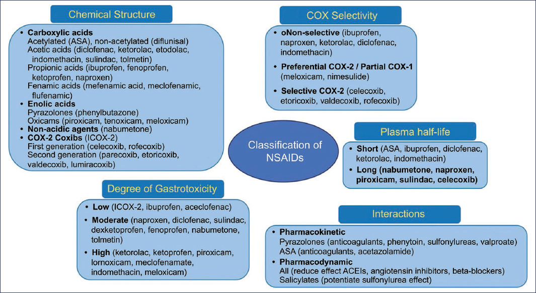
Figure 1. Classification of non-steroidal anti-inflammatory drugs (NSAIDs). ASA: acetylsalicylic acid; ICOX-2: cyclooxygenase 2 inhibitors; ACEIs: angiotensin-converting enzyme inhibitors.
NSAIDs that selectively block COX-2, called “coxibs,” have represented a great advance in the control of inflammatory disorders and those associated with chronic pain. However, despite the fact that due to their mechanism of action, from a theoretical point of view, they should not be associated with gastrotoxicity, they are not exempt from side effects. Some important effects observed in elderly patients are fluid retention, uncontrolled arterial hypertension, heart failure, and even coronary disease.17 Although their tolerability is excellent, even at supratherapeutic doses, short- and long-term endoscopic studies have demonstrated an incidence of gastric or duodenal ulcers of approximately 3-5% (a percentage similar to that observed with placebo), and 1-2% may develop some complication.17 Despite this low risk, selective COX-2 inhibitors significantly reduce the likelihood of developing GI ulcerations and complications compared to classic NSAIDs. With the first generation of selective COX-2 inhibitors, two studies, CLASS (celecoxib) and VIGOR (rofecoxib), which included about 8,000 patients with arthritis, reported a statistically significant reduction of 50% in lesions, compared to NSAIDs that inhibit both COX.17,18 Other more recent studies have supported these findings with new selective COX-2 inhibitors, such as etoricoxib and valdecoxib.19 The use of selective COX-2 inhibitors together with ASA is associated with a rate of ulcers similar to that of classic NSAIDs, and possibly lower than with the coadministration of classic NSAIDs and aspirin. When comparing the frequency of side effects of selective COX-2 inhibitors on the lower digestive tract with that of traditional NSAIDs, selective COX-2 inhibitors showed a lower rate of significant clinical events. Serious complications (hemorrhage, perforation, obstruction, ulceration, or diverticulitis) were 54% less frequent with selective COX-2 inhibitors than with naproxen.19
Mechanisms of protection and healing
The ability of the gastric mucosa to resist damage due to endogenous secretions, such as bile, acid, and pepsin, or to exogenous agents (medications), has been called “mucosal defense” and involves different mechanisms, such as mucus and bicarbonate secretion – regulated by PG synthesis –, turnover and repair of damaged epithelium –which depends on adequate mucosal blood flow–, the hyperemic response as a consequence of an inflammatory process –which traps plasma at sites of damage and creates a microenvironment with a high pH–, as well as the inflammatory response itself, during which chemotactic factors are released with recruitment and migration of inflammatory cells that eliminate damaged tissue, foreign material, and microorganisms, and that promote the formation of granulation tissue and healing.20,21 Once a gastroduodenal mucosal lesion occurs, whether erosion or ulceration, a process of “reformation” of the gastric glandular architecture begins from the infiltration of granulocytes to minimize bacterial translocation. Subsequently, the process of angiogenesis and epithelial migration begins, which is stimulated by various growth factors, such as epidermal growth factor, vascular endothelial growth factor, interleukins 1B and 2, interferon gamma, and adenosine, resulting in increased expression of COX-2 in epithelial monolayers, increased vascular flow at the ulcer margins, proliferation of multiple newly formed vessels, and acceleration in the rate of re-epithelialization.8,20–22 Other external agents are involved in the healing process that can delay the healing of damaged tissue, such as gastric acid, which reduces cell migration and maturation of granulation tissue. Therefore, inhibition of gastric secretion is a key factor in the ulcer healing process. In chronic lesions, the healing process is carried out through granulation tissue formation, re-epithelialization, scarring, and contraction of the ulcer base. The sequence of this phenomenon is shown in table 1.
Table 1. Temporal sequence of development and healing of gastroduodenal ulcer
| Time (days) | Phase | Morphological changes | Modification by medications |
|---|---|---|---|
| 0-3 | Ulcer development | Tissue necrosis, inflammatory infiltrate, granulation tissue | No |
| 3-10 | Early healing | Epithelial cell migration, contraction of ulcer base | Gastric antisecretory agents (PPIs, PCABs) |
| 10-20 | Late healing | Angiogenesis in ulcer bed, granulation tissue remodeling, re-epithelialization of ulcer crater | Rebamipide, sucralfate |
| 20-40 | Reconstruction | Glandular reconstruction, muscular mucosae and propria | No |
| 40-150 | Maturation | Differentiation of specialized cells | No |
|
PPIs: proton pump inhibitors; PCABs: potassium-competitive acid blockers. |
|||
Mechanisms of gastroduodenal damage
Gastroduodenal damage occurs when the deleterious effect of gastric acid exceeds the defense mechanisms. The damage mechanisms produced by NSAIDs can be local or systemic, in correlation with their ability to inhibit PG synthesis, and include the following8,20–22:
- – Inhibition of PG synthesis: this is the main mechanism of gastroduodenal damage. It acts through two forms: locally, by inhibiting mucus and bicarbonate secretion, and systemically, through decreased blood flow and gastric bicarbonate levels, as well as decreased migration of inflammatory cells and release of cell growth factors, fundamental for the tissue repair mechanism.
- – Increased tissue permeability: NSAIDs cause an increase in gastric permeability, which is a direct cytotoxicity mechanism that is not dependent on inhibition of PG synthesis, but appears to be mediated by decreased nitric oxide synthesis, and partly due to decreased bicarbonate secretion secondary to a decrease in blood flow to the gastric mucosa. ASA can break the mucosal barrier without inhibiting PG synthesis, through a mechanism called epithelial toxicity, partly due to the acidic properties of the drug itself.
- – Ionic trapping: in general, acidic NSAIDs are more toxic than those with a neutral pKa. Weak acids are easily absorbed in the gastric mucosa, crossing cell membranes, accessing the neutral intravascular medium, where they ionize, becoming water-soluble substances, thus being “trapped,” achieving high concentrations in the gastric mucosa. At this level, NSAIDs can generate direct effects on intracellular processes, altering cellular permeability, affecting enzymatic activities, allowing back-diffusion of hydrogen ions, karyolysis, uncoupling of oxidative phosphorylation, rupture of intercellular junctions, necrosis, erosions, and hemorrhages.
- – Lipoxygenase activation and increased inflammatory response: inhibition of PG synthesis is associated with simultaneous activation of the lipoxygenase pathway with increased leukotriene synthesis, which causes increased synthesis of tumor necrosis factor alpha and leukotriene B4, as well as upregulation of some cell adhesion molecules. Subsequent events can result in ischemic lesions or damage due to an uncontrolled inflammatory response.
- – Proliferation and apoptosis: the integrity of the mucosa is a balance between cell proliferation and apoptosis. NSAIDs can induce increased apoptosis and cell shedding, with consequent secondary cell proliferation, resulting in epithelial, endothelial, and myofibrillar hyperplasia.
- – Alteration in hemostasis: ASA has a platelet antiaggregant effect, due to inhibition of platelet COX, from irreversible acetylation of COX-1 and COX-2, which prevents the conversion of arachidonic acid to PGH2. Although selective COX-2 inhibitors do not inhibit platelet aggregation, if they are administered together with ASA, the damage of each is potentiated separately, due to blockage of the conversion of arachidonic acid to hydroxyeicosatetraenoic acid with consequent inhibition of the production of gastroprotective epilipoxin A1.20
- – Duodenogastric reflux: active metabolites of some NSAIDs, such as sulindac, can reflux from the small intestine to the stomach, increasing the contact time with the gastric mucosa.
- – Reduction in tissue tensile strength: there are preliminary observations that have associated the use of NSAIDs with decreased tissue tensile strength, although studies are required to confirm this association.
Mechanism of enteric damage
The pathogenesis of NSAID-associated enteropathy is very different from that associated with gastroduodenal damage, and includes an interaction of different mechanisms and factors such as topical effect, COX inhibition, interactions of bacteria and bile acids, and overexpression of pro-inflammatory cytokines8,13,23,24:
- – Topical effect: a topical effect, independent of the inhibitory action on COX that requires direct mucosal contact of the drug on the luminal side, is considered the triggering event in most cases. Once the NSAID is absorbed into the cell, it induces mitochondrial damage through vacuolization, uncoupling of oxidative phosphorylation, disruption of the lipid bilayer of enteral cells, edema, and alteration in electrolyte transport. Naproxen and ASA are two examples of NSAIDs associated with alterations in intestinal permeability.
- – Ileal reabsorption and duodenal recirculation: another important mechanism associated with intestinal ulceration is the passage through enterohepatic circulation of some NSAIDs, particularly carboxylic acids that are conjugated in the liver, excreted into bile, uncoupled by beta-glucuronidases in the small intestine, and subsequently reabsorbed in the terminal ileum, to then be re-secreted into the duodenal lumen, resulting in a double intestinal pass, with potential repetition of damage mechanisms (inhibition of PG synthesis, increased permeability, release of inflammatory mediators, and epithelial damage). Some conditions associated with increased bile flow to the ileum may be associated with increased risk of damage, while measures (experimental or therapeutic) that decrease this flow may reduce the degree of damage.
- – Inhibition of PG synthesis: it has been described that, although there is inhibition of PG synthesis, there is no direct correlation between the degree of suppression and the presence of enteric damage, nor temporal synchronization, so it is not the main pathophysiological factor associated with damage.
- – Dysbiosis-bile acid interaction: animal studies have shown that the administration of NSAIDs (particularly those with enterohepatic recirculation) in germ-free rodents results in significant changes in different types of enteric bacteria, particularly an increase in the number of gram-negative bacteria, which have an increased capacity to deconjugate bile acids and cause epithelial damage. Currently, it is considered that it is an interaction of the intestinal microbiota and bile acids that results in activation of innate immunity, activation of toll-like receptors, and activation of the inflammatory cascade, particularly interleukin-8 and nuclear factor kappa B. Indomethacin is an example of an NSAID associated with cytokine overexpression.
- – Gastrointestinal hypomotility: although it is a secondary mechanism, it has been described that NSAIDs can decrease motility throughout the entire digestive tract and cause from alterations in gastroduodenal accommodation to constipation.
- – Coadministration with other drugs: various medications can induce damage to the intestinal mucosa through different mechanisms involved in the development of damage or alterations in healing mechanisms, including vasoconstriction and ischemia (potassium supplements, contraceptives, methotrexate), cytotoxic damage (oncological treatments including immunotherapy, chemotherapy, or radiotherapy), or interference with mechanisms associated with cell repair (antiplatelet agents, anticoagulants, steroids). The joint administration of any of these drugs with NSAIDs increases the risk of gastrointestinal damage.
Risk factors for development of gastrointestinal damage from NSAIDs
Since only a minority of patients who ingest NSAIDs present GI side effects, and of these a much smaller proportion will develop complications, population groups at higher risk should be identified, in order to design primary preventive strategies or, failing that, carry out secondary prophylaxis. Advanced age (> 60-65 years), male sex, smoking, history of APD, type of NSAID, dose and duration of use, ischemic heart disease, and rheumatological disorders associated with concomitant use of anticoagulants, antiplatelet agents and corticosteroids, and H. pylori infection (Fig. 2), are factors that in epidemiological studies have been associated with a higher RR for developing gastroduodenal damage in NSAID users,8,23–35 while conditions associated with dysbiosis and alterations in enterohepatic recirculation and the flow of bile acids through the small intestine are factors associated with enteric damage8,11,12,23,24:
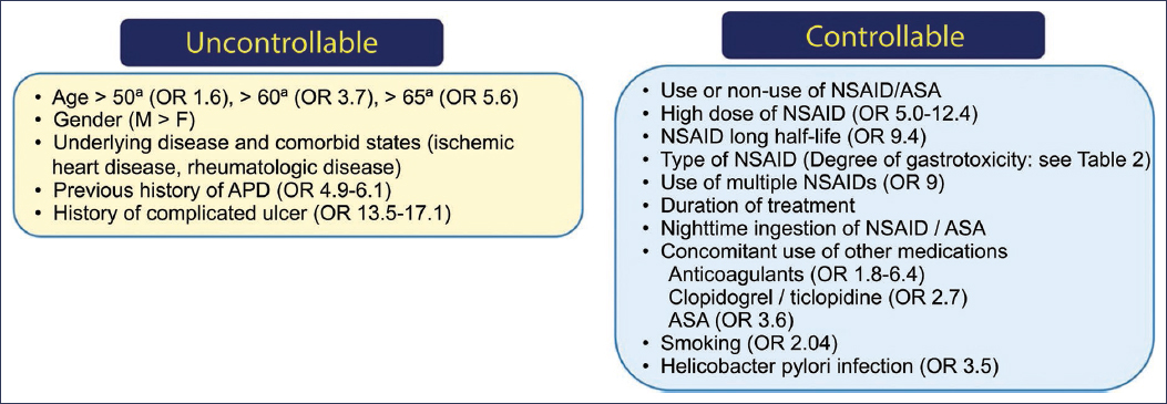
Figure 2. Risk factors for development of gastrotoxicity from non-steroidal anti-inflammatory drugs (NSAIDs). ASA: acetylsalicylic acid; APD: acid-peptic disease; F: female; M: male; OR: odds ratio.
- – Age: adults > 60 years have a higher risk of developing ulcers, regardless of the type of NSAID they use. One study showed that adults > 60 years who took NSAIDs had an RR of 3.7 for developing APD compared to young adults who did not take NSAIDs, and the risk increased to 5.6 if age was > 65 years. When comparing patients with age > 60 years who used NSAIDs with subjects < 60 years without NSAID use, the risk of APD was 13.2 times higher in older patients. It is believed that among the possible causes are changes that occur with age, including decreased PG synthesis, lower GI blood flow, and altered mucus and bicarbonate secretion.
- – History of APD: patients who have had previous APD have a 4.9 times higher risk of developing GI damage when re-exposed to NSAIDs, and the risk is higher when APD has been associated with complications such as bleeding or stenosis, with a risk ranging from 13.5 to 17.1 for the development of new complications. It has been proposed that the risk is higher due to local mucosal changes at sites of previous ulceration.
- – Type of NSAID: NSAIDs have been classically divided into those with high risk, medium risk, and low risk of gastrotoxicity (Table 2). This classification has been derived from the results of several meta-analyses, and among the associated factors are chemical structure, pKa and effect on each COX isoenzyme, dose, half-life, and time of use.25,26
- – Dose and duration of NSAID use: in numerous studies, a linear increase in the risk of ulcer complications have observed a linear increase in the risk of ulcer complications with higher doses of NSAIDs, and although some studies have indicated that selective COX-2 inhibitors at supratherapeutic doses are safe, this group is not free from the risk of developing APD, especially in older adults. An associated risk factor is the patient’s underlying disease: those conditions that require higher doses for symptom control are associated with a higher frequency of complications. For example, 5-10% of patients with rheumatoid arthritis discontinue treatments due to side effects, and about 1.5% of these patients will have a serious complication in the first year of treatment, compared to only 0.73% of patients with degenerative osteoarthritis, a joint disease with a lower degree of inflammation that can be controlled with safer analgesics, such as acetaminophen.25–27
- – Use of anticoagulants: coadministration of anticoagulants and NSAIDs increases the risk of bleeding associated with ulceration (odds ratio [OR]: 3.01) and major bleeding (OR: 2.77), probably due to their antihemostatic properties.28 The use of aspirin also further increases the risk of bleeding in patients taking anticoagulants. Until a few years ago it was thought that coadministration of selective COX-2 inhibitors and anticoagulants did not increase the risk of GI bleeding, but a recent meta-analysis has reported that the risk when coadministered with warfarin is similar to that of other NSAIDs.29
- – Use of corticosteroids: although for many years corticosteroids were considered ulcerogenic, recent evidence has shown that only patients who take NSAIDs together with corticosteroids, especially at high doses (prednisone > 10 mg/day), have a higher risk of bleeding from APD.
- – H. pylori infection: the RR of bleeding from APD associated with H. pylori infection is 1.79, from NSAIDs is 4.85, and from both is 6.13, which suggests an additive effect. Various studies have shown that H. pylori increases the risk of the first episode of ulceration, that eradication reduces the incidence of APD in the general population, especially in patients starting NSAID use, and that eradication reduces the risk of re-bleeding in patients with previous APD.30–35
- – Other factors: there are inconsistent studies about the role of other possible risk factors for APD, such as smoking and alcohol intake, as well as sex and an underlying disease that requires NSAID therapy. Risk factors for enteric damage, unlike gastroduodenal damage, have not been well established, but it has been described that concomitant use of other drugs (gastric antisecretory agents, antibiotics) and predisposing conditions associated with dysbiosis (infections, intestinal surgeries, small intestinal bacterial overgrowth) increase the risk of NSAID enterotoxicity.
Table 2. Estimated relative risks of gastrotoxicity according to NSAID type25,26
| NSAID | Relative risk of gastrotoxicity | 95% CI |
|---|---|---|
| COX-2 inhibitors | 0.8-1.3 | 2.0-5.3 |
| Aceclofenac | 1.4-2.6 | 0.9-4.6 |
| Diclofenac | 2.1-3.1 | 1.6-4.2 |
| Ibuprofen | 2.5-4.1 | 1.8-3.0 |
| Sulindac | 4.2 | 2.8-6.3 |
| Acetylsalicylic acid | ||
| 100 mg/day | 1.8-2.7 | 1.4-3.6 |
| 500 mg/day | 5.6-7.5 | 4.4-9.9 |
| Fenoprofen | 4.3 | 2.8-6.6 |
| Naproxen | 4.0-7.3 | 2.8-11.4 |
| Lornoxicam | 3.5-7.7 | 1.2-24.4 |
| Indomethacin | 3.3-9.0 | 1.7-20.7 |
| Meloxicam | 3.6-9.8 | 1.8-23.8 |
| Ketoprofen | 6.5-8.6 | 2.3-29.2 |
| Tolmetin | 8.5 | 4.5-16.1 |
| Meclofenamate | 8.7 | 4.6-16.4 |
| Piroxicam | 7.2-12.6 | 4.8-20.3 |
| Ketorolac | 8.0-2.7 | 3.4-39.9 |
|
COX-2: cyclooxygenase 2; 95% CI: 95% confidence interval. |
||
Clinical and endoscopic spectrum
The clinical spectrum of gastrointestinal complications of NSAIDs is very heterogeneous, and can involve one or more segments of the digestive tract. The two most affected portions are the stomach and small intestine.36,37 The degree of gastroduodenal mucosal damage ranges from petechiae and subepithelial hemorrhages to erosions, ulcerations, and their complications.35–38 In the small intestine, in addition to the above, the typical lesion is called a diaphragm and involves circumferential mucosal damage, with stenosis and potential obstruction.38–40 Damage progression may vary depending on the type of NSAID. One or two hours after aspirin ingestion, hemorrhages develop in the intact epithelium (petechiae) and superficial ruptures in the mucosa (erosions). With continued use, these changes are reduced due to a process called adaptation. GI damage associated with other NSAIDs develops more slowly, although different factors influence its location, progression, and clinical manifestations, including the type of NSAID, dose, and administration schedule. The most common clinical manifestation is dyspepsia (10-38.5%) and the most frequent endoscopic lesions are duodenal ulcer (45%), gastric ulcer (20-27%), erosions and esophagitis 7%, although it can also manifest as occult GI bleeding or iron deficiency anemia. In the small intestine, it has been described that most lesions are ulcerative, multiple, varioliform, superficial, and with a diameter < 1 cm in 67% of cases; diaphragm disease is observed in less than 5-10% of cases, can affect more than one intestinal segment, and rarely affects the ileocecal valve,40,41 so its presence is considered a surrogate of NSAID damage.42
Treatment
Treatment objectives should be directed toward the phase in which the patient is at the time of medical intervention: primary prophylaxis, treatment of the acute event, management of complications, or secondary prophylaxis, since the pathophysiological mechanisms and degree of damage are different (Fig. 3).
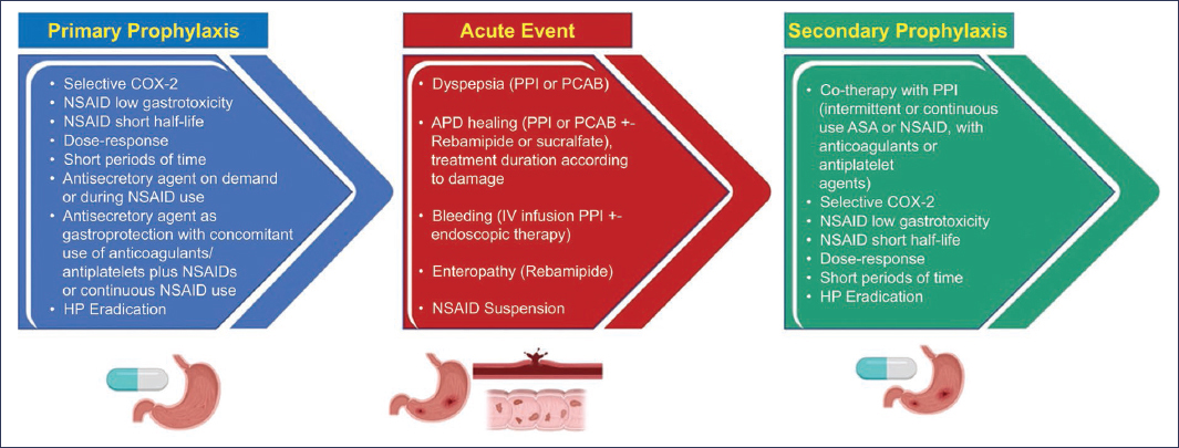
Figure 3. Prevention and therapeutic strategies for gastrotoxicity associated with non-steroidal anti-inflammatory drugs (NSAIDs) (created in BioRender [biorender.com/kasibuk] by Gómez-Escudero et al.12). HP: H. pylori; PPIs: proton pump inhibitors; COX-2I: cyclooxygenase 2 inhibitors; IV: intravenous; PCABs: potassium-competitive acid blockers.
There are several general management strategies, some of which can be applied in more than one phase, which are:
- −Use of NSAIDs with low degree of gastrotoxicity, for short periods of time and according to dose-response.
- −Use of selective COX-2 inhibitors.
- −Locally-acting and tissue-repairing agents (rebamipide, sucralfate).
- −PG agonists (misoprostol).
- −Gastric antisecretory agents (H2 blockers, proton pump inhibitors [PPIs], potassium-competitive antagonists) as gastroprotectors or for healing of gastroduodenal lesions.
- −Combined therapy with NSAIDs and antisecretory agents.
- −Eradication of H. pylori infection.
- −Endoscopic or surgical treatment (or both).8,11,27,36,37
Primary prophylaxis
It refers to medical intervention before the patient ingests any NSAID, in order to prevent the development of symptoms or complications. The first consideration is to select an NSAID with a low degree of gastrotoxicity and short half-life, use it according to dose-response and for short periods of time, choose a COX-2 inhibitor, or, in case of requiring prolonged use, combine it with a gastric antisecretory agent.43–48
USE OF COX-2 INHIBITORS
More than 20 studies have demonstrated a 73-80% reduction in APD when using a COX-2 inhibitor instead of a conventional NSAID.27 One of the first compared ulceration rates at 24 weeks in users of rofecoxib (25 or 50 mg), ibuprofen (2.4 g), or placebo, with rates of 9.6% and 14.7% for rofecoxib doses, 45.8% for ibuprofen, and 9.9% for placebo.43 The VIGOR (Vioxx Gastrointestinal Outcomes Research) study compared the development of important GI effects with rofecoxib (50 mg/day) and with naproxen (1 g/day), with the complication rate being lower with rofecoxib (2.1 per 100 years/patient vs. 4.5 per 100 years/patient).44 Another study compared celecoxib (400 mg/day) with a combination of diclofenac (150 mg/day) and omeprazole (20 mg/day) for 6 months. Although the study concluded that both strategies were similar for preventing bleeding events from APD, the celecoxib group had a lower percentage of lower digestive tract bleeding events (6.2% vs. 10.5%), which suggested that the protective effect of omeprazole is only gastroduodenal and does not prevent NSAID enteropathy.45 Another study compared celecoxib with joint treatment with lansoprazole and naproxen, and obtained the same findings, with the only difference that the coxib was associated with a higher frequency of dyspepsia.46 More recently, etoricoxib at doses of 60 or 90 mg was associated with a lower rate of uncomplicated GI events compared to diclofenac at a dose of 150 mg/day, but there were no differences in complication rates.47 A Cochrane meta-analysis found a lower rate of gastroduodenal ulcers, complications, and treatment suspensions when using COX-2 inhibitors compared to other NSAIDs.48
GASTRIC ANTISECRETORY PLUS NSAID/COX-2 INHIBITOR
Patients at high risk of developing APD when starting treatment with NSAIDs (age > 65 years, history of APD, high doses of NSAIDs, coadministration of ASA, antiplatelet agents, anticoagulants, or steroids) should receive a low dose of a PPI to prevent complications. Evidence comes from nine studies that together report a 54-76% decrease in the risk of developing APD, both in periods < 12 weeks and 12-24 weeks or > 24 weeks.27 The benefit has been reported with most PPIs from the first study (OPPULENT, Omeprazole versus Placebo as Prophylaxis of Ulcers and Erosion from NSAID Treatment), in which 3.6% of the omeprazole group (20 mg/day) developed ulcerations vs. 16.5% of those who received placebo,49 to subsequent studies that showed lower ulceration rates when administering esomeprazole (20 or 40 mg) to NSAID or COX-2 inhibitor users,50,51 or a probability of remaining free of ulcerations with pantoprazole of 95%,52 so this strategy is the current standard of treatment.11,27 Evidence with H2 antagonists is less, but meta-analyses have shown that a single dose is sufficient to prevent duodenal ulcers, but a double dose is required to reduce the risk of gastric ulcers,53 and they are cost-effective in comparative models of strategies that combine H2 antagonists, PPIs, or misoprostol with NSAIDs or selective COX-2 inhibitors, with a 33-68% reduction in the risk of APD, but there are no controlled studies evaluating their long-term efficacy, and they are associated with tachyphylaxis after 4 weeks of use.27
Potassium-competitive acid blockers (PCABs) are the most recent group of gastric antisecretory agents, which have been shown to be non-inferior to PPIs in terms of healing of gastric ulcers,54,55 and although there are no studies yet evaluating their usefulness as gastroprophylaxis for NSAID use, studies in other high-risk groups for APD suggest that they may be useful.56
PROSTAGLANDIN ANALOGS (MISOPROSTOL) WITH OR WITHOUT PPIS PLUS NSAID/COX-2 INHIBITORS
For many years, misoprostol at doses of 400-800 μg/day was used both for primary prophylaxis and for ulcer healing and secondary prophylaxis in NSAID users. The preventive effect has been evaluated in 12 controlled studies, with reductions in the risk of APD of 55-74% compared to the use of NSAIDs and placebo, and with a greater benefit when used for at least 3 months.27,57 A Cochrane meta-analysis concluded that misoprostol was the only drug that reduced the risk of complications associated with ulcers.53 However, due to its mechanism of action, it presents multiple side effects, such as fever, headache, diarrhea, abdominal pain, and increased uterine activity, which are more common with increasing doses, which, when safer drugs are available, limits its use.
REBAMIPIDE
It is a quinolone-derived agent with cytoprotective properties associated with stimulation of PG secretion and gastric mucus, increased blood flow, and decreased inflammatory infiltrate. Administration of rebamipide in healthy volunteers who were given indomethacin or ibuprofen showed a decrease in the risk of gastropathy evaluated by endoscopy,58,59 with a correlation between gastric mucosal flow and absence of antral erosions,59 although a later study did not demonstrate histological protective effect in volunteers who took naproxen.60 A study in chronic NSAID users compared rebamipide (300 mg/day) with misoprostol (600 μg/day) for 12 weeks and found similar rates of gastric ulceration (20.3% vs. 21.9%), but a lower rate of adverse events and fewer treatment suspensions in the rebamipide group.61 An Asian study that evaluated a multicenter database reported that continuous use of rebamipide in new NSAID users for osteoarthritis or low back pain significantly reduced the risk of gastrointestinal bleeding compared to intermittent use.62 Two subsequent meta-analyses reported the superiority of rebamipide vs. placebo against gastroduodenal damage induced by NSAIDs,63 and one of them reported a comparable effect to PPIs in terms of preventing mucosal ruptures and an additive effect to PPIs.64
PRIMARY PROPHYLAXIS OF NSAID ENTEROPATHY
Having a different pathogenesis, the approach with gastric antisecretory agents is usually not useful, and an even higher risk of intestinal damage has been reported, apparently associated with changes in microbial composition.65 There is preliminary evidence in case series or open studies with misoprostol, metronidazole, sulfasalazine, and sucralfate.23 Rebamipide has been evaluated in conditions associated with mucosal ulceration in areas not exposed to gastric acid. In one study, healthy volunteers who received diclofenac for 7 days were assigned to receive rebamipide or placebo, and when evaluated by video capsule endoscopy, a lower rate of damage was observed in the group treated with rebamipide.66 A similar study with diclofenac and PPIs or rebamipide found no differences between groups.67 A subsequent study compared the effect of adding rebamipide or placebo in a group of patients who took ASA and omeprazole, and when evaluating intestinal damage with endoscopic capsule, although no differences were observed in the number of erosions in the jejunum, rebamipide was associated with a significantly lower number of mucosal ruptures in the terminal ileum at 1 and 4 weeks.68 Another similar study found a significant reduction in the number of mucosal ruptures (p = 0.046) and improvement in the Lewis endoscopic score of intestinal damage when comparing rebamipide (200 mg/day) with placebo for 8 weeks, prior administration of ASA for 3 months, with video capsule study before and after starting treatment.69 In a subsequent meta-analysis, rebamipide was superior to placebo in the prevention of intestinal lesions (RR: 2.70; 95% confidence interval: 1.02-7.16).63
An approach aimed at correcting dysbiosis associated with different conditions and that can increase the risk of NSAID damage includes the administration of probiotics, and there is preliminary evidence of the usefulness of some strains such as Lactobacillus casei, VSL#3, and Saccharomyces boulardii.70,71
ACUTE EVENT
Once the patient has developed symptoms or endoscopic manifestations of NSAID gastrotoxicity, medical or endoscopic treatment (or both) is required in order to promote healing of erosions or ulcerations, and to prevent complications derived from them. Two classic studies evaluated the healing rate of APD in patients actively taking NSAIDs: ASTRONAUT (Acid Suppression Trial: Ranitidine versus Omeprazole for NSAID-Associated Ulcer Treatment), which included 541 patients on continuous treatment with NSAIDs and peptic ulcer or > 10 gastric erosions assigned to omeprazole 20 or 40 mg/day or ranitidine 300 mg/day for 4-8 weeks, with an outcome of ulcer healing, reduction to < 5 erosions or resolution of dyspepsia, which showed the superiority of omeprazole over ranitidine (80% and 79% vs. 63%),72 and OMNIUM (Omeprazole versus Misoprostol for NSAID-Induced Ulcer Management), which evaluated 935 patients with the same inclusion criteria as ASTRONAUT and found similar healing rates of gastric ulcer for omeprazole and misoprostol.73 Numerous subsequent studies have documented the efficacy of different PPIs and, recently, of PCABs.
MANAGEMENT OF COMPLICATIONS
With the introduction of PPIs (initially omeprazole, in 1988), and currently with PCABs (vonoprazan, tegoprazan, and fexuprazan), the risk of complications associated with APD has decreased. However, when they do occur, they have high morbidity and mortality, especially bleeding from the digestive tract as a result of erosion of an ulcer into an artery.
INTRAVENOUS PPIS
Intragastric pH affects coagulation mediators, as well as clot dissolution. At pH < 5, peptic activity promotes clot dissolution, but at pH > 6, platelet aggregation is promoted, pepsin is inhibited, and hemostasis is optimized. This is the reason why antisecretory drugs are administered during an acute bleeding event. H2 antagonists cause only a modest increase in intragastric pH, and can generate tolerance after 7-10 days of use; additionally, meta-analyses have not shown a decrease in re-bleeding, surgery, and mortality rates in patients with peptic ulcer and bleeding treated with intravenous H2 antagonists.74 PPIs have been shown to be useful for stabilizing the clot and reducing the risk of re-bleeding. Administration of omeprazole, 80 mg intravenous bolus, followed by an infusion of 8 mg/h in patients with bleeding peptic ulcer, significantly decreased the rate of re-bleeding at 3 and 30 days, so it is now the standard for initial management before endoscopy.75 A meta-analysis that analyzed 21 controlled studies concluded that PPIs reduced re-bleeding rates by 54% and the need for surgery by 41%.76 Another meta-analysis subdivided studies with intravenous PPIs at high or low doses, and found that at a constant infusion (6 mg/h) the PPI was associated with a significant decrease in re-bleeding, surgical event, and mortality rates.77 A Cochrane meta-analysis concluded that, despite the heterogeneity of studies, PPIs were associated with a modest reduction in transfusion requirements and hospital stay.78
PCAB
PCABs block acid secretion more rapidly, achieve peak plasma concentration in 1 hour, are not affected by the previous level of acid secretion or proton pump activity, have a longer-lasting action effect, and are not affected by food. Being a relatively new drug group, evidence of its usefulness is just beginning to emerge.55 A retrospective study compared the effect of prior administration of pantoprazole (80 mg intravenously in single dose) or tegoprazan (50 mg orally in single dose) on the degree of ulcerations and severity of bleeding, and reported that the proportion of ulcerations in stage IIa or higher according to the Forrest classification was significantly lower in the group premedicated with PCAB, as was the frequency of need for therapeutic endoscopic intervention and the rate of re-bleeding.79
Secondary prophylaxis
It refers to medical intervention in a patient who has previously taken NSAIDs or ASA, has developed GI symptoms or lesions, successfully treated, but requires chronic use, so a different therapeutic maneuver or a drug that reduces the risk of developing symptoms or complications again is necessary.
SUSPENSION OR CHANGE OF NSAIDS
This simple maneuver decreases recurrence rates from 50-60% to 0-2%, can be associated with healing of damage up to 95% of cases, and reduces recurrence from 40% to 9%,36 so clinical guidelines recommend suspension or, when not possible, using a COX-2 inhibitor at the lowest dose that controls symptoms, and if it is chronically, add a gastric antisecretory agent.11,37,80
GASTRIC ANTISECRETORY AGENTS
There is evidence of the usefulness of both PPIs and PCABs as secondary prophylaxis. The SCUR (Scandinavian Collaborative Ulcer Recurrence) study evaluated 175 patients with a history of APD who restarted NSAIDs and demonstrated that omeprazole (20 mg/day for 3 months) was superior to placebo (treatment failure rate 24.7% vs. 50%) in preventing the development of new ulcers.81 The ASTRONAUT and OMNIUM studies had a second maintenance phase in which it was evaluated whether patients remained in clinical and endoscopic remission after 6 months on medical treatment. In ASTRONAUT, continuous use of omeprazole (20 mg/day) was associated with remission in 72% of patients, compared to only 59% with ranitidine (300 mg/day).72 In OMNIUM, omeprazole (20 mg/day) was superior to misoprostol (400 μg/day), with success rates of 61% vs. 48%, and with a lower rate of adverse events.73 Several studies have evaluated the effectiveness of PPIs to prevent recurrence of APD in ASA users. In one of them, omeprazole (40 mg/day for 14 days) significantly decreased the degree of damage and the number of gastroduodenal erosions compared to placebo.82 This strategy has demonstrated its usefulness in patients who require chronic use of ASA or other antiplatelet agents for cardiovascular problems, and who have previously had APD.83,84 Two recent studies with similar design analyzed the effect of vonoprazan, a PCAB, and reported that it was not inferior to lansoprazole in preventing the recurrence of ulcerations at 12 and 24 weeks associated with continuous use of NSAIDs85 or ASA86.
REBAMIPIDE
It has been reported that chronic use of PPIs can exacerbate intestinal damage associated with NSAIDs, and there is preliminary evidence of enteric protection with rebamipide, although it is still insufficient.87
ERADICATION OF H. PYLORI
Current evidence indicates that H. pylori infection increases the risk of APD in chronic NSAID users, apparently due to synergistic damage, with an RR of between 2.7 and 61,88,89 as well as recurrence rates when re-exposed to NSAIDs, and that eradication treatment decreases this risk, including lower risk of developing ulcers and complications,90–94 so current guidelines for dyspepsia, APD, and H. pylori, both national and international, recommend looking for it and eradicating it in patients who are going to start long-term treatment with NSAIDs or ASA, in addition to the usual indications such as active APD and uninvestigated dyspepsia.11,27,37,95–98
Conclusions
- – NSAIDs are the most widely used drugs worldwide for their ability to reduce symptoms associated with inflammatory processes, especially in the older adult population. ASA also has platelet antiaggregant action.
- – NSAIDs exert their anti-inflammatory and analgesic effect by inhibiting PG synthesis, so they induce side effects in different organs, and the digestive tract can be affected in 15-30% of cases. Selective COX-2 inhibitors are associated with lower risk of gastroduodenal damage (2-5%).
- – The GI adverse events of NSAIDs affect any organ of the digestive system. Although the main sites are the stomach and duodenum, they can frequently cause intestinal damage, and include all forms of APD and their complications.
- – There are different treatment and prevention strategies at different levels of exposure: primary prophylaxis, acute event, complications, and secondary prophylaxis.
- – For primary prophylaxis, the use of a COX-2 inhibitor or a low gastrotoxicity NSAID is recommended, for short periods of time and at the lowest therapeutic dose, together with a gastric antisecretory agent during the treatment period. Misoprostol is an alternative, but can be associated with diarrhea and increased uterine activity. Rebamipide is useful in cases of intestinal toxicity.
- – During an acute event, a PPI or PCAB is recommended for healing of ulcers and erosions, or in case of bleeding, clot stabilization and endoscopic treatment if necessary.
- – For secondary prophylaxis, suspension of the NSAID if possible, or using COX-2 inhibitors or low gastrotoxicity NSAIDs at the lowest possible therapeutic dose, together with a PPI or PCAB, is better than misoprostol for preventing new damage in patients who need to continue taking NSAIDs. There is preliminary evidence of the usefulness of rebamipide for intestinal toxicity.
- – There are fewer studies evaluating these strategies in ASA users, but current evidence suggests that the same stage-based strategies are applicable to primary or secondary prevention and to damage associated with ASA.
- – H. pylori infection appears to have a synergistic effect in NSAID users, so evidence suggests that eradication before starting treatment prevents damage, complications, and recurrences.
Funding
The author declares not having received funding for this study.
Conflicts of interest
The author declares having been a speaker for Carnot, Prometis Pharma, and Siegfried-Rhein laboratories.
Ethical considerations
Protection of people and animals. The author declares that no experiments were performed on humans or animals for this research.
Confidentiality, informed consent, and ethical approval. The study does not involve personal data, medical records, or human biological samples, so it does not require ethical approval. SAGER guidelines do not apply.
Declaration on the use of artificial intelligence. The author declares that no type of generative artificial intelligence was used for the writing or creation of content of this manuscript.
